


Rules
If you aren't familiar with phasmophobia, it is more like a case study.
A patient presents himself, at different stages in the clinical setup, they will give you one piece of information.
The piece of information can either be a textbook "evidence" for a disease, or a hidden "trait" of that disease.
Your mission is to guess the disease in as little evidence as possible!
Warning: Some images may be graphic, and are based on photographs taken (hopefully consensually) of people who have had the diseases. Please view respectfully.
ant. Diseases

Bacterial Keratitis
- Can have AC reaction and uveitis in recruiting defense cells.
- Hypopyon may occur
- localised redness in hyperemia
- pain, tearing, photophobia, with sticky purulent discharge
- vision loss or poor va
- Lesions are usually focal and defined, central, and large (>2mm). Typically green-yellow colour
- Dense stromal infiltrates with overlying epithelial ulcer

Adenoviral Keratitis
- Very infectious and can survive very long on surfaces, due to the strength of naked capsid viruses
- Characteristic nummular opacities
- Starts unilaterally, and progresses to bilateral, with red watery eyes, swelling lids
- Sometimes lymphadenopathy and photophobia
- Photophobia implies corneal involvement
- Self limiting
- You typically do not want patients with adenoviral keratitis in the clinic, being able to shut it down completely

Acanthamoeba Keratitis
- 5x more likely in contact lens wearers.
- Chance of obtaining if CL cleaned with water, or interactions with swimming pools and hard water.
- Trauma can lead to this as well
- Late stage can cause epithelial breakdown, making it very rough.
- Widespread deep infiltration,
- Usually starting peripherally and migrating centrally
- Rough epithelium, perineural (nerve following) infiltrates, stromal infiltrates
- Wessely ring potentially for immune defence, and hypopyon
- Identified through cysts, and 18sRNA

Sterile Keratitis
- Not as severe as infectious ones
- Usually peripheral to mid-periphery lesions/infiltrates
- fairly small in size (0.1-2mm) with circular ulcers
- Basically no AC reaction, and no oedema.
- Vision typically not affected, and very little photophobia
- CLPU is an example of contact lens version

HSV Epithelial Keratitis
- The "Great Masquerader" or The Mimic
- Can cause retinal necrosis, but I have not done posterior diseases yet
- HSV-1 dwells in the trigeminal nerve (CNV) for reactivation
- Can form shingles around the ocular adnexa
- Tearing, slight blur, photophobia and slight red eye are all common for viruses
- Forms a distinct ulcer lined by raised dendritic cells. This creates a terminal bulb which when stained by fluorescin is easily detected
- Application of steroids may lead to geographhic ulcers. Worsening may lead to corneal anesthesia, or neutrophic keratitis, which is potentially blinding.

HSV Non-necrotising Stromal Keratitis
- Common in recurrent HSV
- central zone opacification with oedema, and keratic precipitates, primarily composed of WBC on posterior corneal surface.
- The usual viral symptoms like photophobia, blur, tearing, pain occur
- Neovascularisation also occurs, along with an epithelial iron line (brownish line)
- Usually intact epithelium, but chronic recurrence

HSV Necrotising Stromal Keratitis
- More rare, more severe
- Can lead to perforation, which should be checked with the Seidel test
- Can be caused by direct viral infection, or secondary immunological reaction to the viral antigens

HZV Keratitis
- The zoster virus dwells in the ophthalmic division of CNV 20x more than the other divisions.
- Respects midline, and so is usually unilateral, along with the shingles.
- Rash will appear on forehead, and tip of the nose
- Shingles will yield burning sensation, and fevers and chills
- Preauricular lymphadenopathy is common
- Zoster Sine Herpete may occur, in which there is no skin involvement
- Unlike HSV, has a characteristic lightning lesion and is self-limiting.
- Presence of AC reaction, flare, nummular keratitis, disciform keratitis, scleral atrophy, neurotrophic keratitis and persistent pains

Fungal Keratitis
- Very similar to bacterial keratitis
- Ulcer is less green-yellow, and more grey-white
- Lesions are not well defined, and instead have feathered edges, along with various satellite lesions.
- Has hyphae lines extending into the cornea
- Gradual onset with a foreign body sensation
- Detected through the hyphae lines on confocal microscopy, KOH staining and Sabourand's agar.
- Stronger and more severe in tropical areas

Phlyctenulosis
- Bacterial hypersensitivity reaction
- Caused by reaction to bacterial antigens, and can usually second tuberculosis
- Usually unilateral, and is characterised by marching phlyctenules (raised white bump) with a leash of vessels
- Neovascularisation occurs from the conjunctiva, and is localised
- Intense photophobia, decreased VA, tearing and blepharospasms
- Treated with topical corticosteroids and antibiotics if affecting cornea
- Conjunctival version is usually quickly self-resolving. Usually spontaneously resolves in 2-3 weeks

Marginal Keratitis
- Bacterial hypersensitivity reaction, usually to s. aureus.
- May second blepharitis
- Photophobia, tearing, irritation, red eye but usually not a big reduction in VA
- Recurrences can be common
- Bilateral most of the time.
- Marked by paralimbal anterior stromal infiltrate.
- Maybe multiple or coalesce.
- Usually at the 4 or 7 oclock position for lesions, and there is a clear area between the limbus and lesions.
- Localised hyperemia in conjunctiva
- Spontaneous resolution in 1-2 weeks, but lid hygiene can help (similar to blepharitis treatment)

Interstitial Keratitis
- Bacterial hypersensitivity reaction
- Could be of infectious origin such as viruses, helminth parasite, tuberculosis, syphilis, or autoimmune disease.
- IMO similar to phlyctenulosis, but pain is usually less severe
- Photophobia, blurry vision, ocular aching, tearing
- Neovascularisation grows from the limbus and is deep-stromal, creating the salmon patch pattern seen
- Often the neovascularisation is a ghost vessel, being vessels with no blood flow or content.
- Treated with topical corticosteroids

Aniridia
- Rare congenital condition, affecting 2.5/10000
- Typically seen at birth with dilated pupils
- Associated nystagmus, photophobia and glaucoma.
- VA usually worse than 20/100
- Can be associated with WAGR syndrome, which is Wilm's tumour, Aniridia, Genitourinary problems and Range of developmental issues

HSV Linear Endotheliitis
- A type of severe endotheliitis.
- Oedema occurs peripherally, and there is an underlying advancing line arranged as keratic precipitates.
- Oedema takes on a sectoral or circumferential shape
- Requires aggressive treatment with topical and systemic antivirals, and corticosteroids.

HSV Disciform Endotheliitis
- Minimal pain, significant photophobia, blurred vision
- Significant deep oedema with intact epithelium. Uveitis present, higher IOP
- Can involve ant. chamber and involved
- Keratic precipitates, cells, flare
- Central epithelial and stromal oedema
- Wesseley Ring precipitates can also exist.

Congenital Glaucoma
- Corneal enlargement (buphthalmos)
- IOP increased and is usually bilateral but is asymmetrical
- Characteristic blue eye appearance
- Characteristic Haab Striae due to cornea being under stress
- Loss of corneal transparency, sensory deprivation amblyopia, poor visual outcome, even with IOP control

CLARE
- Suddenly wakes from sleep by symptoms or not noticed until after waking.
- Moderate pain, redness and tearing along with photophobia
- May be due to bacteria such as pseudomonas aeruginosa
- Usually no treatment is required as there is no real colonisation.

CLPU
- Inflammatory reaction of the cornea
- Characterised by focal excavation of epithelium, infiltration and necrosis of anterior stroma, leaving a scar (green dot).
- Differential diagnosis with marginal keratitis, and bacterial keratitis.
- Hyperemia is localised to around the ulcer.
- Related to CL wear, and is less severe as a result
- Unlike marginal keratitis, may not be parallel with the limbus

Optical Onchocerciasis
- Caused by the nemotode helmnith parasite (round-worm) from the black fly
- Onchocerca Volvulus
- The microfilariae migrates through subcutaneous tissue and skin to the eye tissues.
- Here, due to a 6-30 month lifespan, it will die, causing inflammatory responses
- Causes pruritus and high itching/scratching which could be intermittent or unremitting.
- People blinded have a life expectency 10 year post onset

Thygeson's Superficial Punctate Keratitis
- An unremitting disease with frequent recurrence (every 6-8W) for many years
- Unknown etiology. The image shows Thygeson's accompanied by a dendritic lesion.
- Irritation, burning, photophobia and tearing.
- Characteristic multiple small grey epithelial lesions. Requires differential diagnosis from adenoviral keratitis.
- Usually bilateral, but asymmetrical.
- Treatment with lubrication therapy, mild topical corticosteroids. No response to antiviral

EBMD or Cogan's Microcystic Dystrophy
- 2nd decade onset
- Characterised by maps, dots and fingerprint shaped epithelium basement membrane protrusions.
- Maps: BM protrusion
- Fingerprint: parallel rows of thickened BM
- Dots: Deep epithelial cells trapped under BM, becoming necrotic and forming cysts
- Thickening caused by deposition of fibrillary material between BM and Bowman's
- No hemidesmosomes leads to recurrent erosions.
- May be associated with morning pain and blurring, due to corneal erosions exposing the nerves. Leaves a stabbing sensation.
- Treat using contact lenses

Meesmann's Epithelial Dystrophy
- AD on 12q13 or 17q12
- 1st decade onset
- Tiny cysts within epithelium of uniform size, varied density.
- Mainly central, may reach limbus
- Glare, irritation and light sensitivity all mild, and minimal effect on acuity.
- Intraepithelial cysts may burst, causing great pain

Reis-Bückler's ES Dystrophy
- AD on 5q31, affecting TGFBI gene
- 1st - 2nd decade onset
- Granular deposits in Bowman's membrane.
- Greyish white fine round and polygonal opacities. Rod like and reticular.
- Is progressive, creates a pattern of irregular bands of collagen.
- Eye irritation and pain, increased tearing, photophobia and poor VA. Quite aggressive
- May require excimer laser keratectomy
- Fibrous tissue replaces Bowman's layer

Theil-Behnke's ES Dystrophy
- AD on 10q24
- 1st decade onset
- Similar to Reis-Bückler's but with more honey-comb like appearance
- Curly collagen fibres in Bowman's layer
- Usually less aggressive than RB and has a calmer impact on vision

Granular ES Dystrophy Type 1
- AD on 5q31
- 1st decade onset
- Is progressive. Starts off with small dots that resemble snowflakes/crystals (salt and pepper)
- Crumb like white deposits in the stroma can coalesce in later stages, impacting vision
- Usually spares the periphery
- Deposits are fairly defined compared to macular, but not as defined compared to Type 2
- When stained with Masson trichrome, only hyaline deposits present

Avellino's Dystrophy
- AD on 5q31
- Onset in the 2nd decade. Also known as Granular Dystrophy Type 2
- Usually has opacities that are similar to rings or discs.
- Some resemblance with lattice dystrophy due to linear opacities in the deeper regions.
- Stroma is usually clearer than Type 1, and histologically has hyaline and amyloid deposits after Masson trichrome and Congo red staining
- As deposits increase, stroma will get more hazy
- Photophobia, glare, will later

Lattice T1 Dystrophy
- AD on 5q31
- Onset in the 1st decade
- Usually has fine linear spider-web like branches, characterised by strange linear opacities
- It is reflective, giving the anterior stroma a glossy appearance
- Recurrent erosions + photophobia.
- Since erosions affect the epithelium and reveal the nerves, will also have morning pain.
- Later on, becomes very hazy in the stroma
- More central, so spares periphery

Lattice T3 Dystrophy
- AD for T3(a), AR for T3 on 5q31
- Onset in the 4th - 6th decade
- Unlike T1, there is minimal intervening haze, though the lines coalesce into thicker rope-like lattices.
- This occurs from limbus to limbus with no sparing of periphery
- Enhanced by trauma, may be asymmetric or unilateral for a while
- Treated via PKP or DALK

Macular Dystrophy
- AR on 16q22
- Massive similarities with Granular, however, the stroma is hazy whereas granular does not have that cloudiness
- Progressive vision loss but usually painless
- Entire cornea has hazy patches, and thinning of stroma occurs
- Usually not too recurrent after PKP or DALK
- Histologically will show glycosaminoglycans (GAGs) in the corneal stroma, and mucopolysaccharide deposits

Schnyder's Dystrophy
- Also known as Crystalline Corneal Dystrophy
- 3rd Decade Onset
- AD on 1p36
- The surface is very shiny and crystal like, due to subepithelial crystalline opacities.
- This decreases corneal sensitivity, leading to decreased photopic vision.
- Occurs with deposits of cholesterol and phospholipids in the cornea stroma
- Treated via excimer laser keratectomy

Fuch's Endothelial Dystrophy
- Most common endothelial dystrophy, and 3x more common in females, as well as those over the age of 50
- May have family history, but usually sporadic and is slowly progressive
- Photophobia, blurred vision.
- If you see the potholes in endothelium, most likely Fuch's
- Treated by 5% NaCl due to removing water. PKP or bandage contact lenses
Stage 1:
* Guttata (Hassal-Henle bodies) appear due to thickening of Descemet's membrane and deposition of BM and collagen --> beaten metal appearance
Stage 2:
* Loss of endothelial junctions, leading to loss of active ionic transport, causing oedema and blurry vision. --> worse in morning
Stage 3:
* Bullous keratopathy, which can be painful when bursts. Folds in Descemet's membrane and vision loss due to oedema.

Posterior Polymorphus Dystrophy
- Rare, but reportedly AD
- Asymptotic, and asymmetric onset
- Treatment usually not needed.
- Subtle vesicular endothelial patterns may become confluent, and these can become multilayered to display epithelium like characteristics.
- Thus the abnormal epithelium like proliferation will create band-like vesicular lesions
- Stromal and epithelial oedema seen in rare cases.

Keratoconus
- Central corneal ectasia, usually spares the periphery
- Often comes with Vogt's Striae which are folds in Descemet's membrane due to stress and pressure
- Often comes with Fleischer Rings, which is an iron line deposit, similar to non-necrotising HSV stromal keratitis
- Munson Sign: Contour forms a V-shape
- Rizzutsi Sign: The steeper the contour, the greater the focus of a pentorch light on the iris
- Retinoscopy: Irregular scissor reflect and multiple images
- Refraction: Unclear endpoint, sudden increase in astigmatism, good pinhole VA but poor VA through refraction. Polyporia
- Chauleux Oil Drop Reflex: During ophthalmoscopy, will have a gradient present
- Keratometry and Topography: Bends towards inferior periphery
- Pachymetry: Thickness of cornea varies over cone diameter
Incipient Stage:
* K = 45D, mires are slightly distorted
* No Munson Sign
Early Stage:
* K < 50D, mire distortion
* Slight Munson Sign
Advanced Stage:
* K = 50-56D, mire distortion
* Vogt Striae, scarring, Fleischer ring, Munson sign
Severe Stage:
* K > 56D + tissue changes + munson
* Acute corneal hydrops
- Treated by soft contacts and spectacles in early stage, rigid gas permeable contacts in all stages
- Collagen Cross-linking using riboflarin (vit. B)
- INTACTS: Ring segments that push periphery.
- PKP or DALK

Pellucid Marginal Degeneration
- Similar to keratoconus, except with very noticeable thinning at the 4 or 8 o'clock position depending on your view (image is 4 o'clock)
- Peripheral cornea ectasia, spares the central
- Often comes with Mooren's ulcer, which is a crescent shaped ulcer
- Bulges just above the thinning

Keratoglobus
- Limbus-to-limbus ectasia
- Thinner at edges
- Can be congenital and adult-onset
- Associated with collagen synthesis disorders, and can have blue sclera
- Can have neovascularisation
- Majority of the cornea is now fairly steep

Meretoja's Syndrome
- Lattice Dystrophy Type 2 [false dystrophy]
- 3rd Decade Onset
- A systemic disease caused by amyloid buildup in the trigeminal nerve and corneal stroma.
- Lines are random and scattered, but usually more delicate.
- Is associated with a puppet face due to nerve palsy and inability to express facial expressions
- Lax, itchy skin
- Treated by PKP or DALK

Punctal Stenosis
- Progressive constriction of the puncta.
- Alleviated by anesthetics to dilate and irrigate. However, effect is not permanent.
- Punctoplasty can also be an option, which involves snipping the punctum.
* dilate first, then cut vertically on the ampulla, and horizontally along the canaliculus.
* create a flap and remove the base

Chronic Canaliculitis
- Usually in lower canaliculi
- Rare infection caused by a bacteria, leading to unilateral epiphora and chronic mucopurulent discharge
- May cause conjunctivitis, and treated by foreign body removal or topical antibiotics
- Pouting of puncta, medial conjunctival injection and medial lid oedema.
- Tearing occurring, mild tenderness over nasal aspect of eyelid.

Dacryocystitis
- Infection of the lacrimal sac. Can be acute and chronic. -> epiphora and unilateral conjunctivitis
- Is more medial compared to orbital cellulirits, and can be distinguishing factor
Acute:
* Caused by H. Influenzae, S. Aureus, Strep Pyogenes.
* Oedema, redness, warmth and can worsen into preseptal and orbital cellulitis
* Secondary to PANDO and SANDO
* Systemic antibiotic and warm compresses. May require hospital admission and referral.
* Don't dilate
Chronic:
* Painless swelling but requires DCR

Dacryoadenitis
- Rare inflammation of lacrimal gland
- Unilateral pain, redness and swelling of lateral 1/3 of upper eyelid (due to location of gland)
- Many tearing and discharge
- Upper temporal chemosis
- Chemosis has an S-shape shown in image
- Restricted EOM, and treatment varies with suspected cause
Bilateral Dacryoadenitis:
* Usually a systemic disease
Unilateral Dacryoadenitis:
* Penetrating injury, or spread of bacterial conjunctivitis.

Dacryops
- Round ductal cystic lesions, arising from palpebral lacrimal gland.
- When transilluminated, glows
- Protrudes into superior fornix and is frequently bilateral
- Treated by sucking it out (aspiration)

Pinguecula
- Yellow-white lesion on bulbar conjunctiva which is typically adjacent to limbus, and typically nasal
- Thinning and thickening of epithelium, keratinisation and loss of goblet cells
- Has tissue vascularisation, degeneration of elastic tissue and collagen hyalinisation
- A response to UVA,B, drying or irritation and usually treatment is not required
- Differential diagnosis against pterygium
- Associated with wind, dryness

Pterygium
- Also related to UVA,B
- Whitish triangular fibrovascular subepithelial ingrowth from bulbar conjunctival tissues, with Stocker's line (iron line)
- Degenerated bulbar conjunctiva
- Also adjacent to the limbus, and the apex grows towards cornea
- More common in equatorial regions due to heat and lack of sunglasses. Thus treated with sunglasses or tear supplements. Bowman's layer breakdown.
- Smoking reduces pterygium
- Growth can induce iregular astigmatism, cause redness and irritation, and gritty eye.
- Stromal overgrowth of fibroblasts and BV with inflammatory cells.
Type 1: <2mm onto cornea, and asymptomatic, but accompanied by inflammation
Type 2: 4mm onto cornea, recurrent post surgery, tear film disturbance and astigmatism
Type 3: >4mm onto cornea, affects vision greatly and recurrent lesions

Conjunctival Papilloma
- A sessile papilloma usually in middle aged patients
- Common on bulbar or juxtalimbal conjunctiva, with a history of UVA,B exposure
- Treated by excision
- Can be OSSN (ocular surface squamous neoplasia)
- Can lead to SCC (squamous cell carcinoma) --> has prominent feeder vessels, and persistent pain. Papilloma is typically asymptomatic, but with slight irritation. Also less defined

Primary Acquired Melanosis
- Can be epithelial (PEM) or acquired (PAM)
PEM:
* Pigmentation of basal epithelium due to extra melanin
* Areas of flat patchy pigmentation scattered about conjunctiva, with more at limbus.
* Usually bilateral, benign and non-progressive
* Can be more intense around vessels, and have Axenfeld Loop
PAM:
* Pre-malignant. Usually later in life and can be with or without cellular atypia.
* With = high risk with all layers affected
* Without = low risk with basal layer affected
* Areas of flat patchy pigmentation
* Usually unilateral and can progress into a malignant atypia form.

Conjunctival Haemangioma
- Small sized tumour that is usually asymptomatic or causes mild ocular irritation.
- Composed of endothelial cells of vessels, increased number of abnormal vessels filled with blood
- Benign, slowly progressive, and bright red patches have ill-defined margins
- Typically rounded, nodular, polypoid or lobulated, and can grow to be fairly large.
- Treated through excision, electrolysis and thermocoagulation.

Acute Bacterial Conjunctivitis
- Caused by gram(+) bacteria like s. aureus, h. influenzae
- Acute onset, red and gritty eye, burning and discharge
- Bilateral (1-2 day lag), sticky lids, diffuse conjunctival injection, intense papillary reaction over tarsal plate
- Self-resolving after 5 days, and more red in fornices
- Eyelid oedema, can have profuse purulent discharge, superior corneal ulceration

Gonococcal Keratoconjunctivitis
- Caused by the bacteria N. Gonorrhoeae usually from genitourinary tract infection, but can reach the eye.
- Hyperacute, profuse, purulent discharge, hyperemia and chemosis.
- Swelling may occur, and pseudomembrane formation.
- If very severe, perforation and endophthalmitis (infection of ant. chamber) can occur
- Perforation tested with Seidel Test
- If progression, requires urgent systemic and topical antibiotics, and hospitalisation.

Adult Chlamydial Keratoconjunctivitis
- Caused by serotypes D->K of Chlamydia Trachomatis g(-)
- Requires host cell for replication
- Transmission as STI or eye-to-eye spread
Signs:
- Bilateral redness, and mucopurulent discharge
- Large follicles, mostly in inferior fornix, but can involve superior.
- Preauricular lymphadenopathy
- Will have superficial punctate keratitis occasionally, and chronic cases may develop less prominent follicles, and instead papillae
Treatment:
- May have variable peripheral keratitis and treated through topical erythromycin, tetracycline, systemic azithromycin
- However, topical antibiotics are insufficient alone, and only provide relief.
- Effective treatment requires systemic antibiotics.
- Additionally, symptoms may take weeks to settle

Trachoma
- Caused by serotypes A, B, Ba and is the classic trachoma
- person-to-person transmission or via vectors like house flies
Active Phase:
* Conjunctival inflammation (follicle and papillae)
* Limbal follicles, and watery discharge
* Keratitis
Cicatricial Phase:
* Tarsal conjunctival scarring especially upper. Can be linear, stellate and then confluent
* Distortion of eyelid and trichiasis
* Secondary trichiasis and entropion leads to corneal scarring and blindness
* Resolved limbal follicles result in gaps in corneal pannus (Herbert's Pits)
Trachomatous Inflammation - Follicular (TF)
- Presence of five or more follicles in the upper tarsal conjunctiva, which are at least 0.5 mm in diameter.
Trachomatous Inflammation - Intense (TI)
- Pronounced inflammatory thickening of tarsal conjunctiva, obscuring more than half the deep tarsal vessels
Trachomatous Scarring (TS)
- Presence of scarring in tarsal conjunctiva. Bands o white lines/sheets are visible and glisten.
Trachomatous Trichiasis (TT)
- At least one eyelash rubs on the eyeball. Should be removed
Corneal Opacity (CO)
- Easily visible corneal opacity. Pupil margin is blurred, and VA will be worse than 6/18 or 0.3 logMAR.

Neonatal Conjunctivitis
-Ophthalmia Neonatorum
Bilateral conjunctival inflammation developing within 1st month of life
- Infection is from the maternal parent
- Purulent discharge, conjunctival swelling, redness and follicles
Gonococcal:
* 1st week onset with aggressive treatment to prevent perforation
HSV:
* 1-2 week onset with watery discharge
Staphylococcus and Others:
* end of 1st week
Chlamydial:
* Most common, and 1-3 week onset.

Adenoviral Keratoconjunctivitis
- Will usually have preauricular lymph node palpation (lymphadenopathy)
- Pseudomembrane formation, hyperemia, subconjunctival haemorrhages, chemosis
Acute Non-specific Follicular Conjunctivitis
* Mild symptoms like follicles and redness, with no corneal involvement and self-limiting in 7-10 days.
Pharnygoconjunctival Fever
* Serotype 1, 3, 7
* Affects children typically, with upper respiratory tract infection and is bilateral.
* 30% manifestation of mild keratitis
* Burning, tearing, photophobia, fever
Epidemic Keratoconjunctivitis
* Serotype 8, 19, 37
* Highly contagious, but no sore throat or fever
* 80% manifestation of severe keratitis
* Unilateral usually, and 2-3 weeks to self-limit
Chronic Adenoviral Conjunctivitis
* rare, but very persistent. Chronic non-specific follicular papillary lesions occur
* History of acute conjunctivitis prior
* Periods of tearing, redness, photophobia

Molluscum Contagiosum Conjunctivitis
- Caused by the DNA poxvirus, which is transmitted via contact in children, and sexually in adults.
- Chronic unilateral irritation and mild discharge
- Is usually self limiting
Eyelid Infection:
- Usually painless with watering and photophobia
- Skin shows painless waxy, umbilicated nodules or papule.
- 2-3 diameter nodules has central depression with no visible inflammation
- Ipsilateral hyperaemic conjunctivitis with follicles, pannus in long standing cases
- Superficial keratitis
- If immunosuppressed, can occur in multiple or confluent regions
- Nodules can appear cheesy and white, consisting of infected degenerate cells.
- 6-12 month self-resolving
- Can be removed by laser, cryotherapy
- Generally left alone unless very close to lid margin

Allergic Conjunctivitis
Acute Rhino:
* Allergic reaction to allergen. IgE-mediated response
* 20% of population suffer, most develop in children, and second group develops post pubescent allergies
* Sudden swelling, itching, sneezing, lid oedema and erythema
* Conjunctival chemosis
Seasonal (SAC):
* Onset during spring and summer as hayfever
* Most frequent allergies include tree and grass pollen with geographic variation
Perennial (PAC):
* Through year, and allergies include dust mite, fungal, animal, genetic predisposition
* Less common and less severe than SAC
- Managed through antihistamines, mast cell stabilisers and usually not steroids.

Vernal Keratoconjunctivitis
- Bilateral recurrent disorder involving IgE and cell-mediated immunity
- Increased IgE in tears and serum
- Conjunctival epithelium contains eosinophils and mast cells
- Primarily impacts boys of age 7-10, especially in warmer climates. 95% remit whilst the other 5% develop into atopic
- Seasonal variation present
- Characterised by papillae palepebral conjunctiva, limbal gelatinous hyperplasia and corneal involvement.
- Manifests with intense itching, tearing, redness, photophobia, burns, thick mucous discharge
Palpebral Disease:
* Papillary response (left image) that is so rough it scratches the cornea, especially upper
* Mucous can manifest between giant papillae
Limbal Disease:
* Hyperemic, oedematous and thickened.
* Gelatinous papillae at limbus consists of small elevated lesions, consisting of desquamated epithelial cells and eosinophils, known as Horner-Trantas Dots (right image)
Corneal Disease:
* Usually upper 1/3, with punctate epithelial keratophy.
* Scratching and mucous may induce peripheral superior neovascularisation
* Plaque can form to create a smooth shield ulcer
- Topically treated with mast cell stabilisers, antihistamines and low dose steroids
- Systemically treated with immunosuppressants or antihistamines
- May involve surgical removement of plaques

Atopic Keratoconjunctivitis
- Rare bilateral disease that is chronic and persistent.
- Hx of atopic eczema or dermatitis
- Corneal thinning occurs, leading to sensitivity to lots of allergies.
- Atopic implies genetic predisposition towards immediate hypersensitivity, thus positive family Hx.
- Type I and IV hypersensitivity reactions, and contributes to inflammatory changes of conjunctiva and cornea
- During exacerbation, increased IgE in tears and serum. Increase B-cells, but depressed T-cells
- Similar to VKC but more and unremitting.
- Eyelids are red, thickened, fissured lids with chronic staphylococcus blepharitis and madarosis.
Conjunctiva Specific:
* Micropapillary conjunctivitis upper and lower tarsus, fornix.
* Giant papillae may develop over time.
* Cicatricial conjunctivitis with inferior shortening of conjunctiva, with symblepharon and caruncle keratinisation.
Corneal Involvement: (Atopic Keratopathy)
* Punctate lesions now in the lower 1/3 opposite to VKC
* Plaque formation may occur, along with peripheral neovascularisation
* Risk of infections.
- Treatment is similar to VKC, but less effective and so prolonged treatment needed
- Systemic immunosuppressants, oral antihistamines.
- Surgery to remove plaques and amniotic membranes.

Mucous Membrane Pemphigoid (MMP)
- Chronic subepithelial blistering disease which affects the mucous membranes and skin, forming scars.
- 75% with oral involvement also have conjunctival MMP
- 25% with skin involvement also have conjunctival MMP
- The conjunctival MMP (cMMP) is also known as ocular cicatricial pemphigoid, and only affects the eye, without the mouth or other skin.
- More prevalent in females by over double, impacting those around 70 y/o.
- Early presentation <60Y indicative of prognostic signs
Conjunctival Signs:
* Persistent conjunctivitis with subepithelial vesicles that could rupture and cause ulcers
* Subepithelial fibrosis, symblepharon, entropion and trichiasis
* Goblet cell loss, reducing mucin secretion.
* Severe dry eye, leading to corneal scarring, ulcers and neovascularisation
* Has an insidious onset, chronic slowly progression, exacerbations. Redness, irritation, burning and fevers. Conjunctival injection and necrosis
Lids and Cornea:
* Flat plica, keratinised caruncle
* Blepharitis and keratisation of lid margin.
* Anklyoblepharon or adhesion of upper and lower lids
* Limbal cell failure, causing corneal neovascularisation and secondary infection
* Dry eye, exposure keratopathy
* Keratinisation and conjunctivalisation of corneal surface -> increases risk of secondary bacterial keratitis
* Diffuse hyperaemia, pesudomembrane formation and symblepharon.
- Image shows both symblepharon and keratinisation of cornea
- Systemic treatment needs physician control
- Steroids can be used, but not for long term as it cannot fully contain disease
- Chronic treatment with dapsone, which is a surface antibiotic.
Stage 1: Chronic conjunctivitis with mild corneal involvement
Stage 2: Cicatrization with conjunctival shrinkage and foreshortening of fornices
Stage 3: Presence of symblepharon, distortion of lashes.
Stage 4: Ankyloblepharon and several corneal involvement

Stevens-Johnson Syndrome (SJS)
- Also known as Toxic-Epidermal Necrolysis (TEN)
- Rare disease, and prognosis for OCP/cMMP
- Ocular involvement is less common for TEN
- More common in males, around age 25
- SJS affects < 10% of body SA, whilst TEN affects >30%
- Affects skin and mucous membranes by separating the epidermis from the dermis layer.
- Triggered by medication, infections, genetic predisposition
- Fever, fatigue, sore throat -> painful ulcers and lesions -> rash develops on body and epithelial sloughing occur -> 20-25% mortality
Skin Rashes:
* Diffuse skin rashes and mouth mucous ulceration
Acute Ocular Disease:
* Crusty eyelid, transient self-limiting papillary conjunctivitis and pseudomembraneous conjunctivitis
* In late stage: scarring of upper tarsal plate
* Poster lid margin, conjunctival scarring and symblepharon
* Dry eyes due to goblet cell destruction and fibrosis of lacrimal duct
* Corneal keratinisation and keratopathy can be secondary to cicatricial entropion, aberrant lashes, infections and ulcers or neovascularisation
Limbal Stem Cell Failure:
* Persistent epithelial defect and corneal neovascularisation
* Conjunctivalisation, keratinisation, dry age, exposure keratopathy.
Systemic Therapy (control active disease):
* Acute controls: steroids, cyclophosphamide, antibiotics
* Chronic controls: immunomodulation via dapsone, azathioprine, mycophenolate
Local Therapy (control inflammation):
* Ocular lubricants to control complications for rehabilitation, epilation and haptic lenses to prevent trichiasis
* Keratopathy controlled through retinoic acid, limbal stem cell transplant, lamella graft or keratoprothesis

Superior Limbic Keratoconjunctivitis (SLK)
- Chronic inflammatory disorder which are usually bilateral in nature, affecting upper limbus and thus associated superior bulbar conjunctivitis.
- Worse in the mornings, gets better throughout the day, and worsens at night again.
- 50% keratoconjunctivitis sicca (drying)
- related to TED or can be idiopathic
- Burning, phhotophobia, mucoid discharge, FB sensation and mechanical rubbing all possible
- Superior corneal punctate staining
- Loose superior bulbar conjunctiva, leading to papillary inflammation
- Papillary hypertrophy of superior tarsal conjunctiva

Ligneous Conjunctivitis
- Very rare disorder but will occur at any age.
- Due to a plasminogen defect, leading to build up of fibrin
- Recurrent bilateral firm fibrin-rich lesions on tarsal plate
- Gradual onset and may be covered by yellow-white thick mucoid discharge
- Histology shows eosinophils and coagulated fibrin on conjunctival surface
- Tissue remodelling may occur

Trichiasis
- Abnormal misdirected growth of eyelashes
- Commonly acquired secondary to chronic blepharitis , tarsal plate or conjunctival scarring from Trachoma or SJS. Can also be due to HZV ophtalmicus
- Aging and entropion
- Pain relief from pulling eyelid away
- Treated either by tackling underlying causes, solving corneal epithelial defects or removing aberrant eyelashes
- Can use electrolysis, cryotherapy, laser or surgery to destroy hair follicles
- Follicles are anatomically normal, and eyelid is typically neither inverted nor everted

Distichiasis
- Rare and is when eyelashes begin to grow from the Meibomian Glands. Related to trichiasis
- Can be congenital, with the germ cell which differentiates into MB instead grows into a pilosebaceous unit
- Can be also acquired if Meibomian glands transform into hair follicles or pilosebaceous units. This is from metaplasia due to intense conjunctival inflammation
- Acquisition may be associated with chemical injury, OCP and SJS
- Treatment tends to be through surgical excision, cryotherapy or lasers
- Can also apply cryotherapy on posterior lamella to kill lashes, and reappose to allow healing

Madarosis
- Local disease like basal cell carcinoma.
- Is the general loss of lashes
- Can be related to endocrine disorders
- Infections like leprosy, HIV, syphilis
- Skin related issues, eyelash removal, trauma
- Trauma may be burns, radiation, eyelid tattoos or extensions.

Poliosis
- Eyelash whitening
Ocular:
* Chronic anterior blepharitis
* Sympathetic ophthalmia
* Uveitits
Systemic:
* Vogt-Koyanagi-Harada Syndrome. Attacks melanocytes
* Vitiligo
* Expect IgE

Benign Cysts
Cyst of Moll:
* Lower left
* Due to serous aqueous production, will be more translucent
Cyst of Zeiss:
* More sebaceous, and so will be more opaque and oily-white looking
- Superficial and Deep cysts may also occur
Milia:
* Milk cyst, and occurs in crops usually due to sun damage. Caused by occlusion of pilosebaceous follicles or sweat pores

Comedones
- Black heads
- Plug of sebaceous matter, with blackened mass of epithelial debris

Papilloma
Squamous Cell Papilloma:
* Benign lesion, with fibrovascular connective tissue covered by irregular squamous epithelium.
* Flesh coloured, and can either be sessile (right image), or pedunculated (stalk like)
* Has a raspberry like surface
Basal Cell Papilloma:
* Discrete greasy brown plaques or seborrhoeic keratosis. Warts
* Grows slowly with age and very common in the elderly, with proliferation into squamous layers and keratin filled cysts
* Has a pasted on appearance as a flat brown lesion (left image)
* Usually on middle lower lid

Naevi and Naevus Flammeus
Intradermal:
* Most common, elevated, non-pigmented and benign (top right)
Junctional:
* Usually flat, well-circumscribed and uniform brown. Low malignant potential (bottom left)
Compound:
* Both, and usually brown pigmented, with low potential (top left)
Naevus Flammeus:
* Otherwise known as port-wine stain
* Rare-congenital subcutaneous lesions
* Due to ectatic vessels in papilla demis
* Ocularly related to Ipsilateral Glaucoma
* Demarcated soft pink-patches. Is segmental and usually unilateral.
* Does not blotch with pressure, and redness increases with age

Ectropion
- Outward turning of lower lid margin, away from globe
- Epiphora + redness of lid margin, palpebral conjunctiva becomes visible. Puncta not in apposition to globe.
- Pinch test would cause punctum to be displaced laterally.
Involutionary:
* Most commonly affects lower lid of eldery px, and can cause chronic conjunctival inflammation and thickening.
* Laxity of lids, canthal tendons, and poor retraction problems due to degeneration of fibres and elastic
Cicatricial:
* Due to scarring or contraction of skin, which pulls lid from globe.
* Caused by burns, trauma, dermatitis, SJS, surgery
Mechanical:
* Mechanical lid eversion usually caused by lump/tumour
* remove cause if possible, and correct loose lid.
- General treatment can be temporary, through tear substitution, lid taping, or tarsorrhaphy (sew eyelid together)
- Surgical through canthoplasty, scarred lids skin graft, wedge resection

Entropion
- Inwards turning of lower lid margin
- Congenital or involutionary (age-related)
- Scarring via trauma, or trachoma
- Ocular irritation, redness, watery/mucus discharge
- Lash turn-in can lead to corneal abrasion. Is thus recognised as a common cause of eyelash misdirection
- Observable flipping with strong lid blinking due to orbicularis oculi spasms, and so you can ask patient to blink hard
Involutionary:
* Affects lower lid more than upper since the upper lid has a wider and more stable tarsus
* Horizontal lid and canthal tendon laxity or stretching.
* Preseptal eyelid also overrides into the pretarsal orbicularis
* Leads to great irritation, redness and excess tearing. Long term can lead to ulceration and pannus
Cicatricial:
* Scarring of conjunctiva pulls lid margin towards globe, and can either impact upper or lower
* Protection of cornea can be done by removing the inward turning lashes
* Treated with bandage CL. Mucous membrane grafts can also replace scarred conjunctiva.

Anterior Blepharitis
- Inflammatory reaction to exotoxins release by s. aureus and s. epidermis.
- Long term effects leads to lid ulceration and scarring
- Crusting/sticking of eyelid in the morning
- Burning, irritation, FB sensation, tearing and photophobia.
- Redness + oedema, telangiectasia (dilation of small vessels)
- Punctate staining adjacent to lid margin
- Collarettes and surf debris on eyelashes, and eyelash matting of lashes and madarosis
Staphylococcal (Top Left):
* Recurrent, and will have scales around lashes, and hyperaemia of anterior lid margin.
* Madarosis, poliosis, trichiasis + phlyctenulosis
* Lid scarring, notching and ulceration.
Angular (Bottom Left):
* Due to bacterial overpopulation, leading to red and scaly skin at canthus
Seborrhoeic (Right):
* Associated with dandruff
* Oily, scaly and shiny skin with soft debris and potential meibomian gland
- The more severe, the more sticking, burning, irritation, eyelash debris, redness and thickness of oedema.
- Treat with warm compresses, lid scrubs
- Topic antibiotic and steroids, but only for max 2 weeks

Posterior Blepharitis
- Also known as Meibomian Gland Dysfunction (MGD)
- Chronic diffuse abnormality of Meibomian Glands. Duct is obstructed, leading to buildup of sebaceous oils, causing alternation of tear film and eye irritation.
- Similar symptoms to ant. blepharitis
- Inspissated or plugged MG secretions, causing oil globules at the MG opening (left image)
- Pressure on lid margin will ooze fluid out.
- Expressability measured using a device to consistently quantify pressure to grade MGD
- Associated with dry eye and poor tear film stability in 30-50% of the time. This can lead to foamy tear films (right image)
- Notching of mid margin also occurs
Grade 1:
* Minimal altered expressibility and secretion quality + no symptoms
Grade 2:
* Mildly altered expressibility and secretion quality + mild symptoms and minimal staining
Grade 3:
* Severely altered and marked symptoms with high staining -> punctate corneal erosions.
Plus Disease:
* co-existing or accompanying disorders
* Exacerbated inflammatory ocular surface disease

Demodex Blepharitis
- A type of posterior blepharitis caused by demodex mites
- forms cylindrical dandruff (shown) at root of eyelashes, and eyelashes also misdirected (shown)
- Main differentiating factor from staph. aureus
(Chronic Blepharitis - Tangent):
- In general, lid hygiene, warm compresses, tear substitutes, antibiotics are important.
- In addition, there are also nutritional supplies to help MGD, and lubricating eye drops to combat tear film instability and dry eyes.

Chalazion
- Chronic granulomatous inflammatory lesion caused by blockage of meibomian gland orifice.
- Retained sebaceous secretion from meibomian, or other sebaceous glands like Zeis.
- Gradually enlarging lid nodule
- Usually painless
- Occurs at all ages
- May affect vision if large and pressing on cornea
- Lid eversion shows roundish nodule within tarsal plate
- Usually self-limiting or can be surgically removed.

Phthiriasis Palpebrarum
- Lice on Lids
- Image on left shows an example of lice mimicking eyelid eczema (shown in allergic eyelid reactions)
- Can be caused by pediculosis capitis (head lice)
- Pediculosis corporis (abdomen lice)
- Pediculosis pubis (crabs) -> STI and if found in children as phthiriasis palpebrarum, suspect child abuse.
- Itching, FB sensation and conjunctival injection
- Remove lice and smooth with ointment for 7-10 days. Re-examine after a week.

Allergic Eyelid Reactions
Acute Allergic Oedema:
* Sudden onset, painless, red
* Insect bites, hives and angioedema.
* Unilateral or bilateral, and chemosis is present. But is self limiting.
* Like most allergic reactions, treated with mast-cell stabilisers, antihistamines, and cold compresses
Contact Dermatitis:
* Inflammation of skin from repeat allergen exposure.
* Can be due to topical medication or preservative. Sensitised on first exposure, and T-cell hypersenstivity next time
* Tearing, painless, itching, red eye, uni/bilateral
* Treatment: remove allergen, use drops without preservatives to prevent allergic reaction.
* Antihistamines, cold compresses
Atopic Dermatitis (Eczema - shown)
* Very common allergic inflammation of skin. Idiopathic and often associated with hay fever.
* Chronic itching, scratching, thickening of eyelid skin
* Treated via low dose steroids, moisturised skin and infection treatment

Acute Horedeolum
Internal:
* Staphylococcus aureus infection of the Meibomian glands
* Tender painful swelling within the tarsal plate, and can discharge through skin and conjunctiva.
* On the outside, resembles dacryocystitis and dacryoadenitis, but when turned in, reveals the internal infection.
External: (stye)
* Staphylococcus infection at lash follicle and associated gland of Moll/Zeis. Usually Zeis
* Can be distinguished on outside of eyelid
- Lid hygiene, warm compresses and massages for 10 minutes 4x a day.
- Don't squeeze or stab
- Topical or oral antibiotics

Impetigo
- Also known as school sores
- Caused by s. aureus and s. pyogenes
- Redenning of skin and bullae blisters form
- Forms gold-yellow crusts.
- Painful erythematous macules rapidly develop into thin-walled blisters.
- May be fever, malaise. Treatment is usually topical and sometimes oral antibiotics.

Erysipelas
- St Anthony's Fire
- Acute subcutaneous spreading cellulitis
- Is diffuse. Surface layer of skin is infected (upper dermis)
- Diabetes, obesity and alcohhol abuse can predispose this
- Caused by s. pyogenes usually through minor trauma
- Well defined, indurated, tender red plaques. This distinguishes it from other cellulitis
- Treated by oral antibiotics
- Firey burning sensation, chill and fevers

Necrotising Fasciitis
- Very rare infection
- Called flesh-eating disease, and caused by s. aureus and s. pyogenes (more often)
- Affects limbs, trunks and preorbital regions, affecting soft tissues. + post-operative regions.
- Rapidly progressive necrosis, redness and selling in pre-orbital regions
- Secondary tissue death occurs due to thrombosis
- may cause bilateral lid necrosis affecting elderly or debilitating.
- Periocular is rare, redness and oedema followed by large bullae and black discolouration.
- Treat through early surgical debridement, systemic antibiotics.

Preseptal Cellulitis
- Bacterial infection of tissue that is anterior to the orbital septum, including fat and dermis
- High risk of orbital development
- s. aureus, epidermis, pyogenes.
- Due to skin trauma (lower lid as shown), lacerations, insect bites
- May be due to local infections, like acute hordeolum, dacrocystitis, dacroadenitis
- May be due to upper respiratory tract infections (rarely)
- Risk increased with diabetes mellitus
- Acute onset of swelling, redness, tender lids, fever, malaise and irritability
- Erythema of skin, lid oedema, ptosis and pyrexia (fever > 38)

Orbital Cellulitis
- Bacterial infection of tissue that is posterior to the orbital septum, and thus within orbit.
- Is a severe and vision-life threatening emergency
- Caused by sinus-related spread, preseptal cellulitis, local spread from dacrocystitis or dental causes.
- Can also be a haemotaogenous spread, or post surgical
- s. pneumonae, s. aureus, s. pyogenes, h. influenzae
- Suddenly, onset of unilateral swelling of conjunctiva and lids. Pain in ocular movement.
- Blurred vision and reduced VA + diplopia
- Fever (pyrexia), severe malaise
- Proptosis, restriction of EO motility, and pain with movement
- RAPD and abnormal pupillary response
- Painful ophthalmoplegia and ON swelling which can lead to dysfunction
- Immediate hospitalisation and emergency, with systemic antibiotics, ancillary tests and surgery if antibiotic resistance.

HSV Reactivation
- Reactivation from CNV, and has non-specific facial and lid tingling within 24 hours.
- After 24 hours, eyelid and preorbital vesicles.
- Eyelid and preorbital vesicles on lid margin. Breaks down over 48 hours
- Usually confined to a single dermatome. Randomised distribution that doesn't respect the midline like HZO
- Follicular conjunctivitis, discharge, swelling and HSV keratitis may follow with dendritic ulcers
- According to textbook, papillary conjunctivitis
- Self-limiting over 6-8 days. Very severe cases occur with eczema.
- Oral and topical antivirals needed, and antibiotics to prevent secondary treatment.

Herpes Zoster Ophthalmicus
- Latency in V1 of CNV
- Of V1, VZV often resides in the portions of ganglion that supply upper lid and nasociliary branch.
- 50-70% suffer visual morbidities with increasing severity in 5-8th decade of life.
- Hutchingson's Sign: Skin lesions in inner corner of eye or tip of the nose due to respect of midline.
- Rashes on cheek if V2 affected
- Pain and altered sensation of forehead on 1 said, and general malaise.
- Rash on forehead, and progresses from vesicles to pustules and then crusting
- Red base
- Can cause skin lesions, ocular lesions like keratitis, episcleritis and scleritis
- Neurological complications like nerve palsy and optic neuritis.
- Post-herpetic neuralgia or chronic severe pain
- Treated with acyclovir oral/topical antivirals. Topical corticosteroids if stromal keratitis occurs. Tear supplements

Orbital Mucormycosis
- Rare opportunistic fungal infection mucoraceae
- Typically in Px with diabetic ketoacidosis or immunosuppression
- Aggressive and often fatal.
- Infection spread to contiguous sinus and subsequently to orbit and brain
- Destroys roof of mouth as well
- Blood is invaded by hyphae resulting in occlusive vasculitis with orbital tissue necrosis

Thyroid Eye Disease (TED)
- A consequence of Graves' Disease, which is a form of hyperthyroidism, or the excess production of Thyroxine and Triiodothyronine (T4 and T3, but more T3)
- Graves' Disease tells include goitre, hand tremor, warm sweaty fingers, enlarged fingernails, muscle weakness, swealing, heat intolerance, fatigue, nervousness. Treated through radioactive Iodine-123, partial surgery, or anti-thyroid drugs
- influences middle aged adults, 3-5x more often in women
- Always bilateral, but often asymmetrical. Multiple EOMS involved, yet usually inferior and medial rectus.
- Progressive for 3-5 years before stabilising.
- IgG anti-body mediated.
- Shown by CT scan, EOMs are enlarged due to infiltration by T-cells and fibroblasts
- Cellular infiltration of interstitial tissues, and orbital fat and connective tissues are proliferated.
Active Inflammation:
* Eyes red and sore, can last a few years
* Remission within 3 years usually
* 10% Px develop serious long-term ocular complication
Quiescent Stage:
* Eyes white
* Painless motility defect still present
* Severity may range from being a nuisance to blindness (optic neuropathy)
- Symptoms include grittiness, photophobia, lacrimation
- Decreased VA and diplopia
- Decreased colour perception due to compression of ON and thus also VF defects
- Retrobulbar discomfort (something painful behind eye)
General Treatment:
Manage Exposure:
- Lubricants, moisture shields, punctal occlusions, eyelid taping.
- This will prevent dry eye, keep tears for as long as possible.
- Head elevation when sleeping
Manage Diplopia:
- Prism Rx, strabismus surgery, botulinum toxin
Manage Compressive Neuropathy:
- Surgical decompression, and removal of one or more orbital wall.
Manage Systemic Disease:
- NO SMOKING
- Refer GP and endocrinologist and comanage
- Systemic steroids and radiotherapy carefully

TED - Pt. 2
Soft Tissue Involvement:
* Preorbital and lid swelling
* Conjunctival hyperemia indicates disease activity
* Chemosis. Conjunctival swelling may overflow over eyelids
Eyelid Retraction:
* Retraction of upper and lower eyelid occur in 50% Px
* Normal upper eyelid lies 2mm below limbus
* Sclera now present, and cosmetic problems
* Contraction of levator by fibrosis
* Fibrosis of inferior rectus may similarly induce retraction of low eyelid. May also have Muller muscle overstimulation.
* Characteristic staring appearance (shown in image)
Proptosis:
* TED is the most common cause of both unilateral and bilateral proptosis in adults
* 70% of the time untreatable without surgery
* Prevents lid closure, and so chronic dry eye, corneal ulceration and blindness
* High chance of secondary infection
Optic Neuropathy:
* Serious complication affecting 5% of px. Large and congested EOM compresses the ON and nerve supply, leading to blockage at orbital apex
* May occur in absence of proptosis, but is a severe and preventable visual blindness cause
* Leads to impaired colour vision and VF defects, with a progressive deterioration in VA.
* Atrophy results in severe cases, and management depends on severity
* Usually systemic steroids, radiotherapy, or as a last resort: orbital decompression surgery
Ocular Motility:
* Between 30-50% of TED Px develop eye movement problems
* Restrictive oedema in infiltrative stage and fibrosis during fibrotic stage
* Surgery recommended if diplopia in primary gaze or reading. Won't be a complete fix, but will have good cosmetic results.
* Botulinum toxin injection to relax muscles

Orbital Venous Anomalies (Varicies)
- Abnormally enlarged tortuous vein, artery or lymphatic vessel in the eye.
- Can be congenital, and can cause proptosis which is accentuated by coughing
- Varix may become clotted and bleed
- May be conjunctival or eyelid, where simple coughing will cause vessels to clot up, fill up and push out.

Direct Carotid Cavernous Fistula
- Defect in the intracavernous part of the internal carotid artery
- 75% trauma caused, or spontaneous rupture due to hypertension.
- More common in females
- Pulsatile proptosis can be observed or heard
- Chemosis is present
- Bruit is present, which is the whooshing sound with a stethoscope, which is silenced under pressure.
- Conjunctival vessels are tortuous (refer to next)
- Image shows the carotid artery blockage
- Ancilliary test such as CT and MRI or angiography (shown)
- may spontaneously resolve through thrombosis.
- Surgery requires neurosurgeon referral, but it is not life threatening.
- Lubrication therapy and temporary prism for diplopia

Indirect Carotid Cavernous Fistula
- Caused by the indirect communication between meningeal branches of internal or external carotid and cavernous sinus (dural shunt)
- Slower blood flow, and so clinical features less subtle
- Tortuous veins shown in the image, and above shows the location of high blood pressure and clotting.
- Caused either trough congenital malformation, or through spontaneous rupture via trauma. Straining in those with hypertension.
- Bruit is softer but can still be heard.
- Mild epibulbar injection without chemosis
- Raised IOP, and ophthalmoplegia caused by CNVI palsy, or EOM swelling.

Lymphangioma
- Rare benign non-functional vascular malformations due to haemodynamically isolated non-functional vascular systems.
- Can bleed into lumen and form cyst, and onset is fairly early in 1st decade.
- Can be anterior and posterior, and surgery is fairly tricky

Superficial and Deep Dermoid Cyst
Superficial:
* Present in infancy, with no globe displacement
* superotemporal most common, but occasionally superonasal
* Is freely mobile under the skin, and margins are easily palpable, and cyst can have hair, skin appendages or glands.
* Visualised with CT scans and treated via excision
Deep:
* Presents in adolescence, and leads to non-axial proptosis
* May extend intracrancially is there are bony defects, can cause FB reaction is leakage
* CT scan visualisation and treated via excision

Amblyopia
- Not blur, but low VA and distortion
- Not caused by defects in the eye itself, but is related to neural damage from optic nerve to the visual cortex
- Caused by Strabismus, anisometropia (primarily hyperopic), high astigmatism, isoametropia and form deprivation and suppression.
Strabismus Amblyopia:
* No form deprivation, just reduced vision and conflicting visual inputs
Meridional Amblyopia:
* Deprivation of form in 1 meridian
Anisometropic Amblyopia:
* Deprivation of form in 1 eye, and competition between inputs
Deprivation Amblyopia:
* Deprivation of form, and weak competition due to high bias of one eye
Cortical Damage:
* Fractional Anisotropy is low, thus tissue is less dense
* Mean Diffusivity is high, thus more things are leaking out
* In LGN, layers are poorly defined and under represented. This is due to significantly lower feedback processing.
* V1 Cortex Displays low activation and processing
- Poor Visual Acuity: Not due to blur but due to distortion. Optical corrections can be very effective in treatment.
- Spatial Acuity and Contrast: Amblyopes can't localise vernier acuity. They also have a much lower CSF, especially for higher spatial frequency
- Crowding: Crowding usually doesn't impact vision in the fovea, but it does occur for amblyopic individuals, showing the amblyopic fovea resembles the normal periphery
- Attention Span: Stronger suppression of eye leads to less attention, and thus deeper amblyopia leads to lower attention modulation.
- Motion Perception: Whilst 5-10% of unidirection is required for global motion perception, 30% for amblyopic individuals to see.

Mucocele
- cystic lesion with mucous
- Presents in adults, caused by obstruction of nasal sinus secretions
- Usually due to frontal or ethmoidal sinuses- Slowly expanding lesion which gradually erodes sinus walls.
- Workup requires referral

Encephalocele
- Herniation of intracranial contents through congenital cell defects.
- Meningocele is only dura
- Anterior (fronto-ethmoidal) can cause forward and lateral globe displacement
- Posterior (spheno-orbital) can cause forward and downward globe displacement

Cat-scratch and Self-scratch Disease
- Parinaud Oculoglandular Syndrome or cat-scratch disease is caused by gram(-) Bartonella Henselae from scratches or bites
- Leads to unilateral granulomatous conjunctivitis (shown left) and ipsilateral lymphadenopathy along with fevers.
- Self Limiting
- Factitious Conjunctivitis or self-inflicted conjunctivitis will have various forms, yet can have corneal scarring, subconjunctival haemorrhages, and requires GP and mental health specialist assistance

Lymphanoma
- Salmon patch with smooth surfaced vessels
- Females predominant, and usually in 5-7th decade of life
- 31% with systemic involvement.
- Px with minimal symptoms, leading to average delay of 8 months between clinical onset and diagnosis.
- Conjunctival redness, irritation, excessive tear production
- Non-progressive and non-painful

Corneal Graft Rejection
- Cloudiness occurs
- Can lead to failure, but failure does not lead to rejection
- Antigens on donor button can cause immune response
- Keratic precipitation occurs
- Khodadaust Line: Segmented oedema and endothelial white blood cells adjacent to clear cornea, creating a line (faintly seen)

Sebaceous Cell Carcinoma
- Extra rare sebaceous oil gland cancer
- Yellow/orange painless nodules
- Is linked to Muir-Torre syndrome and UV radiation as well as immunosuppression
- Also Great Masquerader as it mimics benign lesions, like chalazion and chronic blepharitis.
- Atypical presentation of a typical disease is a hint, especially with yellow thickening
- Hallmark is pagetoid spread, as spread of malignant cells to epithelium that appears to be separate from main tumour.
- Very rare, slow growing, affecting elderly, and more so women. Usually arises from meibomian glands, though sometimes from Zeis or elsewhere.
- Histopathologically lobules of cells with pale foamy vacuolated lipid-containing cytoplasm and large hyperchromatic nuclei.
- Mortality is 5-10%, adverse prognostic effects include upper lid involvement, tumour size 10mm or more + duration of symptoms more than 6 months
- Usually on upper eyelid, and can involve both lids on one side (5%)
Yellow Material: Within tumour indicates SGC mostly
Nodular:
* Discrete hard nodule, most commonly within upper tarsal plate. Yellow discolouration due to lipid -> Chalazion mimic
Spreading:
* Infiltrates dermis, causing diffuse thickening of lid margin, often with eyelash distortion and loss. Mistaken for blepharitis

Conjunctival Melanoma
- Can include naevus and melanosis.
- Can be PAM and SAM
- Naevi can develop into melanosis.
- Melanoma found in limbal, bulbar, fornical or palpebral conjunctiva and demonstrated dilated tortuous feeders and intrinsic vessels
- CATCH Melanoma: Children, Age older, Thickness and base greater, Cyst lacking, Haemorrhage and melanoma.

Ptosis (congenital and involutionary)
Congenital
- Usually seen after birth, with poor levator function and fibrotic change.
- Developmental problems with the levator
- Sometimes superior rectus weakness
Involutionary
- Dehiscence, disinsertion or stretching of levator aponeurosis restricts transmission of force from a normal levator muscle
- Worsens throughout the day due to strain on the Müller muscle

3rd Nerve Palsy
- SR, IR, MR, IO and Levator, and PSNS to sphincter and ciliary body.
- Acute onset, but ptosis present, causing diplopia.
- May be accompanied by pain, or mydriasis.
- May be partial or complete. With pupil sparing, it may be ischaemic. Regardless, best to refer to emergency immediately
- Aneurysms can occur near the Circle of Willis, which will cause brain bleeding, and compress 3rd nerve to cause palsy.
Compression Caused:
* Generally pupil involving
* Aneurysms at internal carotid junction
* Brain tumours or raised intracranial pressure
Ischaemia:
* Pupil sparing usually
* Diabetes, hypertension, artheriosclerosis or giant cell arteritis
- Could also be caused by inflammation or trauma.
Tells:
* Affected eye bends laterally (LR innervated by 6th nerve, not 3rd)
* Inability to adduct affected eye, raise affected eye, depress affected eye
* Levator also failing, thus ptosis present

Marcus-Gunn Jaw Winking
- 5% of all congenital ptosis cases
- Usually unilateral.
- Appears to be an aberrant connection between motor branches of 5th nerve.
- Contraction of pteryoid jaw muscles causes an excitation in the 3rd nerve, which innervates the levator ipsilaterally
Tells:
* Retraction or wink of ptotic lid during stimulation of the pterygoid jaw, either through chewing, open mouth, teeth clenching or swallowing

Horner's Syndrome
- Damage or deficiency of the sympathetic pathway.
- Leads to pupillodilator dysfunction.
- Is commonly associated with a triad:
Ptosis: (always present)
* Müller muscle affected (SyNS)
Pupil Miosis:
* Pupil smaller than unaffected side, and dilates slowly in dim light
Anhydrosis: (sweating and flushing)
* 1st Order = ipsilateral body
* 2nd Order = ipsilateral face
* 3rd Order = post-ganglionic
- Observed also with iris heterochromia, where affected iris is lighter, due to innervation effect on tyrosinase in melanocytes
- Congenital or children < 2 years
- Could also be idiopathic
1st Order Defects: (Central)
* Primarily intracranial conditions
* Brain stem lesion, tumours, MS
Encephalitis, meningitis
* Syringomyelia, cyst or cavity of spinal cord
* Spinal cord tumour
* Lateral medullary syndrome
2nd Order Defects: (Preganglionic)
* Affects the transverse thoracic region
* Neuroblastoma of the paravertebral sympathetic chain.
* Cervical rib
* Pancoast tumour (Malignancies involving the lungs)
* Carotid and aortic aneurysms
* Neck lesions/trauma to brachial plexus
3rd Order Defects: (Postganglionic)
* In close proximity to the internal carotid artery
* Cluster headaches
* Nasopharyngeal tumours
* Otitis media
* HZV
* Temporal arteritis
* Cavernous sinus mass, or internal carotid disease
* Carotid cavernous fistula
Tells:
* 0.5-1% apraclonidine is an anti-glaucoma drug with weak adrenergic agonist action. Application reverses anisocoria due to pupil sensitivity to adrenergic reactions
* 10% cocaine prevents norepinephrine uptake at post ganglionic nerve ending.
Due to the lack of NE, the Horner's pupil will not dilate
* 1% hydroxyamphetamine to confirm location of lesion. Preganglionic will reverse anisocoria, postganglionic will not
* Chest X-ray followed by CT scan is required if pulmonary malignancy is suspected, such as from a history of smoking.
* Head CT scan and MRI for possible stroke.
* MRI warranted when there is carotid artery dissection is possible. Painful Horner syndrome.

Myasthenia Gravis
- Autoimmune condition with unknown origin.
- Leads to weakness and fatigability of muscles
- young women in 30s most affected
- Occurs when there is a produced blocking antibody which deposits on neuromusclar junction membrane receptors to prevent the entry of AcH
- Electromyography can confirm muscle fatigue, serum antibodies to detect AcH receptors, and CT for presence of Thymus Tumour
- Treated by anticholinesterases to retain more signal molecules
- Steroids and azathioprine for lower responses
- 2/3 have both ptosis and diplopia
- <10% ptosis alone, <30% diplopia alone
Tells:
* Pertinent negative is that pupils are never involved.
* Ptosis and diplopia may be present, but never pupils.
* Worse at end of the day, least on awakening
* If eyelid manually held in place during upwards gaze, fellow eyelid will show fine oscillatory movements
* Ice pack improves ptosis due to cold improving neuromuscular junctions

Pseudoptosis
- False Ptosis
- Due to enophthalmos, anophthalmos, microphthalmos
- Hypertony (low IOP)
- Hypertropia or hypotropia
- Lid retraction of other eye, leading to perception of ptosis
- Dermatochalasis (excess skin) or brow ptosis

Eyelid Retractions
- Upper eyelid is retracted
- Could be due to TED
- Could be due to neurogenic, such as unopposed levator action, 3rd nerve redirection, jaw-winking
- Could be mechanical, due to scarring, or congenital, due to down syndrome
- Could be idiopathic
- Blepharochalasis -> non-pitting lid oedema
- Eyelid imbrication -> topical over bottom lid
- Varicies -> abnormal vascular formation
- Blepharospasm -> forced involuntary closure of eyelids

Floppy Eyelid Syndrome
- Usually in middle-aged, overweight males
- Redness, irritation, itching, mucous dischage
- Worse upon awakening, and can be both unilateral or bilateral
- Sleep face down, with chronic irritation of everted eyelids
- Complain of erratic sleep and morning headaches
Tells:
* Obstructive sleep apnoea (temporary halt of respiration)

Marcus-Gunn Pupil
- Rapid Afferent Pupil Defect (RAPD)
Damage to the Retina:
* Large, unilateral retinal detachment
* Could be of ischemic cause due to vessel occlusion
Damage to ON:
* Optic neuritis or multiple sclerosis
* Ischemic optic neuropathy, due to stroke or decreased blood supply
Others:
* Severe unilateral amblyopia, cerebrovascular accident or stroke
* Very rarely cataracts
Tells:
* Compare 1 eye to the next. First shine light into one eye. If it constricts, then it is normal
* Quickly swing to other eye. If slight dilation occurs due to weakened response, then RAPD is present
* Can use a neutral density filter equivalent

Argyll-Robertson Pupil
- Bilateral miosed and irregularly shaped pupils.
- Common cause was neurosyphillis, but now more likely sarcoidosis and diabetes.
- Other causes include chronic alcoholism, encephalitis, multiple sclerosis, Lyme disease or HZV.
- Pathophysiology due to a lesion in the dorsal Edinger-Westphal nucleus, interrupting the pretectal oculomotor light reflex fibres.
- Develops at an asymmetrical rate.
Tells:
* Reacts poorly to direct and consensual light, but reacts briskly to near stimuli.
* Indicates near-light dissociation, where potential damage in the pretectal nucleus exists
* This will impact the light reflex, but have an intact near reflex

Adie's Tonic Pupil
- Is a PSNS efferent defect leading to denervation of the afflicted pupil.
- Localised to ciliary ganglion. Unilateral mydriasis and loss of sphincter function
- Can be bilateral
- Typically affects females in the 3rd-4th decade. 3:1 ratio
Tells:
* Mydriasis in affected eye, with sluggish miosis in chronic pupils.
* No response to direct or consensual
* Sluggish response to near, leading to accommodative miosis. This is significantly faster than chronic sluggish light reflex miosis.
* Miosis occurs with 0.125% pilocarpine

Hutchinson and Pharmacological Pupils
Hutchinson's Pupil:
* Unilateral, fixed dilated pupil due to 3rd nerve lesion
* Other pupil contracts and reacts normally.
* Due to severe head trauma, but may also be due to stroke, tumour, abscess, oedema
Pharmacological Pupils:
* Drug-related pupillary reactions
* Mydriatic agents include cycloplegics like tropicamide, cyclopentolate, atropine, hematropine, scopolamine. + phenylephrine
* Miotic agents include pilocarpine

Coloboma
- Failure of optic fissure to close during development.
- Causes absence of ocular tissue, and may be sporadic, hereditary, or associated with chromosomal abnormalities.
- May affect iris, ciliary body, choroid, retina, optic nerve
- In the iris, causes a partial lack of formation of the iris
- Linked to CHARGE syndrome:
Coloboma, Heart defects, Atresia of choanae, Retardation of growth, Genital hypoplasia, Ear malfunction
.jpg)
Persistent Pupillary Membranes
- 30% of patients will have this due to common nature
- Atrophy of PM should be complete by 6MG, and occurs in 95% of normal newborns
- Usually, connected to other parts of the iris.
- If large and not removed, can cause form-deprivation amblyopia.
- Cataracts may also occur with PPM

Axenfeld-Rieger Syndrome
- AD, but rare, with 1 in 200,000
- Has a heterogenous presentation
Post Embryotoxon:
* Anterior displacement of Schwalbe's line. Can be seen (viewable in image)
Iris Abnormalities:
* Hypoplasia
* Corectopia (pupil displacement)
* Polycoria (many pupils)
Systemic Abnormalities:
* Dental (hypodontia)
* Facial bone: broad nasal bridge, maxillary hypoplasia
* Umbilical: redundant periumbilical skin

Iridocorneal Endothelial Syndrome
- Rare disorder of unknown origin, causing angular, corneal and iris damage
- Proliferation of abnormal endothelium with epithelial phenotype. Thus PPED is a differential diagnosis, along with Fuch's ED and Axenfeld-Rieger
- Abnormal endothelial cells, with abnormal clone that takes on epithelial phenotype and characteristics. This migrates onto angle and iris surface.
- Such membrane can then proliferate and enlargen
- Corneal effects: Oedema and decomposition
- Angle effects: Glaucoma
- Iris effects: Corectopia, atrophy, holes, traction and ectropion
Chandler's Syndrome:
* 50% of all ICE. Confined to inner corneal surface
* Diffuse corneal oedema + beaten bronze appearance (similar to Fuch's ED)
* Subtle atrophy of iral stroma and corectopia, and may be accompanied by secondary glaucoma
* Synechiae (sticky iris) forms as endothelium moves over ant. angle.
Essential Progressive Iris Atrophy:
* Unilateral subtle stromal atrophy -> full thickness atrophy
* Includes corectopia, ectropion uveae and stretch-melt holes
* Peripheral ant. synechiae in all Px.
* Endothelial cells and surrounding collagen fibrillar tissue extend from cornea over trabecular meshwork and iris anterior surface. This forms abnormal contractile membrane.
Cogan-Reese Syndrome:
* Iris Naevus Syndrome
* Least common, and has corneal involvement and pigmented lesions on iris. Less severe iris atrophy however
* Shown in image, is distinguished by tan, pedunculated nodules, with different pigmented lesions on the anterior iris
* Loss of normal iris architecture with flat effaced iris surface that may appear darker (heterochromia)
* Associated with glaucoma

Blunt Trauma (pt. 1)
- When force is applied to the eye, the equator will expand, damaging the posterior pole.
- During rebound, the equator will collapse, and the decompression can damage the anterior.
Corneal:
* Abrasion causes epithelial injury, which may be stained. Is very painful, and accompanied by photophobia and tearing in Descemet's
* Acute corneal oedema may be secondary to shock and endothelial dysfunction. This leads to stromal haziness, thickenings and folds.
Anterior Chamber:
* Hyaemphema -> haemorrhage due to ciliary body or iris
* Signs -> Blood is visible in the anterior chamber, usually transient. Monitor IOP
* Symptoms -> Pain, blurred vision, Hx of trauma
* Treat -> use atropine to immobilise iris to reduce further haemorrhage and uveitis. Anti-fibrolytic agents may be used.
* Grade 1: <1/3 filled
* Grade 2: 1/3 - 1/2 filled
* Grade 3: >1/2 filled
* Grade 4: Eight Ball
Uvea:
* Pupils -> severe contusion
* Iridodialysis -> dehiscence (splitting) of iris from ciliary body at its root. D shaped pupil can cause diplopia, glare and will require surgical repair.
* Cyclodialysis -> ciliary body detachment from scleral spur
* Angle recession -> rupture of force of ciliary body, irregular widening of ciliary body. Rise in IOP due to damaged trabecular draining.
* Iridioschsis -> iris stomal layers split
* Ciliary body trauma -> temporal loss of aqueous production. Shock and hypotony (soft eye). Tears in ciliary body
* Secondary Uveitis -> Possible due to trauma

Blunt Trauma (pt. 2)
Lens:
* Cataracts -> pigment imprinting on anterior lens capsule (Vossius Ring) during direct contact, and may have a ruptured lens capsule.
* Sublaxation -> secondary to torn zonular fibres, lens deviates towards intact zonules.
* Dislocation (laxation) -> rare complete rupture of zonules. Lens dislocated towards vitreous.
Globe:
* Very severe blunt trauma
* Check if IOP low compared to fellow eye
* usually anterior, with prolapse of intraocular structures such as lens, iris, ciliary body and vitreous
* posterior ruptures as well, and difficult to detect
Vitreous:
* Posterior vitreous detachment can occur, sometimes with haemorrhages.
* Pigmented cells floating in ant. chamber. This is called Tobacco Dust
Retina and Choroid:
* commotio retinae (shown by whitening)
* choroidal ruptures
Retinal breaks and retinal detachments
Optic Nerve:
* Optic neuropathy, avulsion.

Orbital and Blowout Trauma
- Medial wall is thinnest, yet the floor is the most susceptible to blow out fraction, as it lacks support from the ethmoidal air cells.
- Inferior Rectus muscle can be trapped, since trigeminal nerve may be compressed
- Infraorbital anaesthesia.
- Pain, diplopia, crepitus (nose-blowing with crackling noise), hypoaesthesia (low cheek sensation)
- Bony defects filled with soft tissue and fat from orbit
- Alters support mechanism for EOM, and potentially trapping them.
- Direct muscle damage can result, either from ischaemia or physical
Signs:
- Periocular oedema and variable ecchymosis.
- Subconjunctival haemorrhage, swelling
- Diplopia due to haemorrhage and/or oedema in orbit.
- Defective ocular motility involving abduction and adduction
- Eye sunken in orbit with vertical misalignment
History:
- Time and mechanism of injury, change in appearance
* Immediate loss of vision implies severe retinal damage
* Loss of light perception implies vascular occlusion or ON compression
* Initial good vision implies compression optic neuropathy.
Ecchymosis
* Black eye.
Secondary Injuries:
* Ruptured globe
* Retro-orbital, vitreous haemorrhage
* Hyphaema
* Angle recession and dislocated lens
* Secondary glaucoma
* Retina/choroidal detachment

Penetrating Trauma
- 3x more common in males.
- Commonly caused by occupational, assaults, domestic accidents
- Extent of damage depends on object. Biggest risk is endophthalmitis, creating a risk of infection.
- Inquire about tetanus shots, and when the last one was
Cornea:
* Repair depends on extent of wound, damage to intraocular components and iris involvement
Small wounds:
- Anterior chamber ok, and may not need suturing, but potential management with bandage lens
Medium wounds:
- Suturing usually needed, post-op. bandage lens
Iris:
- If iris involved -> usually requires iris abscission
- If lens damaged -> remove lens due to cataracts, suture laceration.
Sclera:
- Anterior: better prognosis
- Posterior: retinal break associated, and worse prognosis.

Lacerating Trauma
Eyelid Trauma:
* Superficial: parallel to lid margin, no gaping. Suture directly
* Lid Margin: Invariably gape. Carefully suture to avoid notching
* Mild Tissue Loss: Suture + lateral cantholysis, allowing for movement
* Extensive Tissue Loss: Combat with reconstructive effort
* Canalicular: Plumbing needed. Silicone tubing and reconstruction
- Additioanlly, subconjunctival haemorrhages may occur, but usually this is not painful, and is picked up by others
- Conjunctival lacerations can occur, with the conjuncitva fusing to repair.
- Finally, laceration may occur to the cornea, which will require sutures and stitches. Prognosis depends on damage

Superficial Foreign Body
Subtarsal:
- Small particles, seen with lid eversion. Irrigate thoroughly.
- Can use cotton bud with anaesthetics
- Will create linear patterns of scratching over the cornea that can be stained.
Corneal:
- Very common, and creates a lot of irritation.
- Leukocytic infiltration
- If left, risk of secondary infection and corneal ulceration.
- Mild anterior uveitis is common, photophobia, miosis,
- Rust staining if ferrous origin (rust rings can form)
Management:
* irrigate with sterile saline, though usually doesn't work.
* Anaesthetise cornea, remove using foreign body spud, or sterile forceps.
* To remove rust, use alger brush, which removes whole epithelium to prevent rust spread
* Antibiotics to prevent secondary infections, and use cycloplegia if necessary

Intraocular Foreign Body
- Can cause direct or secondary effects via infection, toxic effects etc.
- Can lodge into any structure
- Usually due to fast moving hot metal, which seals wound upon entry.
Management:
* Accurate history, origin of forein body and time.
* entry and exit wounds should be checked, fluorescein, slit lamp, gonioscopy and fundoscopy should be utilised.
* Remove using magnetic removal of ferrous bodies.
* Sclerotomy may cause direct effects or secondary effects via infection or toxic effects
* Forceps removal via pars plana victrectomy, and remove through pars plana or limbus.
Siderosi Bulbi:
* Deposition of iron from foreign body. Can affect lens epithelium, iris, ciliary body, and retina.
* Anterior capsular cataract with radial iron deposits, reddish brown iris and pupillary mydriasis
* If damage to trabecular region, then secondary glaucoma.
Chalcosis Bulbi:
* Foreign body with high copper content usually leads to endophthalmitis and loss of eye.
* Alloy such as brass with low copper can lead to deposition of copper to collagen and BM
* Forms Kayser-Fleischer rings, sunflower cataracts and retinal deposition that may not be degenerative.

Chemical Burn
Urgent Treatment:
- Irrigation and continuously, for at least 10-20 minutes, since permanent damage in seconds.
- If alkaline, irrigate for an hour. Several litres required.
- Evert eyelids to remove trapped matter, and do not contaminate sound eye
- Anaesthetics can help
- Only use saline or water
Acids: Damages ocular surface by denaturing proteins, which create a barrier that limits diffusion.
Alkaline: (2x more common)
* More damage due to fatty acid saponification, disrupting cell walls.
* Diffusion is progressive, and can damage cornea, conjunctiva, lids, trabecular meshwork, iris, lens
1. Destruction of conjunctival cells with fibrosis, creating lid distortion and symblepharon
2. Loss of corneal limbal stem cells, reducing capacity for re-epithelialisation
3. Damage made more exclusive. Due to exaggerated inflammatory and healing response. Stromal melting and thinning occurs
4. Fibrosis of anterior segments. Complicated sequelae with glaucoma and cataracts
Signs:
* Irritated/swollen eyelids
* Blurred vision
* Excruciating pain in both eyes
* Irritated or burnt skin around eyes
Sequelae:
1. Damage
- Necrosis conjunctival and corneal epithelium.
- Disruption/occlusion limbal vasculature -> loss of LESC
- Deeper penetration, breakdown of GAGs, stromal opacification
- Anterior chamber damage to iris and lens
- Ciliary body dmg. hypotony and phthisis (progressive wasting)
2. Healing
- Migration of epithelial cells, and synthesis of new collagen, giving the LESC is not damaged.
Grade 1: Clear cornea, ok limbus, good prognosis
Grade 2: Hazy stroma, iris visible, <1/3 limbal ischaemia
Grade 3: Total loss of corneal epithelium, stroma hazy, iris not visible, 1/3-1/2 limbal ischaemia
Grade 4: Opaque cornea, >1/2 limbal ischaemia, poor prognosis
+ conjunctival epithelial loss, iris change, lens change, IOP
Treatment:
* Grade 1 -> short course topical steroids, cycloplegia and antibiotics
* Grade 2 or worse -> reduce inflammation, promote epithelial regrowth, prevent ulceration.
* Early Surgery to try revascularise limbus, restore LESC and fornices
* Suture tenon's capsule to limbus to re-establish vascularisation
* LESC transplant via allograft or autograft
* Amniotic membrane
* Late surgery including keratoplasty, scleral lens or keratoprothesis

Radiation Damage
- Longer wavelength yields thermal damage.
- Glassblower's cataracts, retinal lesions and regular cataracts
- Short wavelengths yield photokeratitis and snow-blindness
UV-Related Photokeratitis:
* Welder's Flash or Snow Blindness
* UV damages epithelium, causing severe pain, and poor vision
* Lasts few days before re-epithelialisation
* Stromal damage depends on duration.
Glassblower's Cataracts:
* Intensely heated glass/metal emits IR radiation
* Chronic exposure for 10-15 years can cause this premature cataracts.
Thermal Damage:
* Accidental burn with scalding water. Examine cornea and conjunctiva for burns
* Like lice, note possibility of intent or child abuse

Scleral Decolouration
Focal:
Scleral Hyaline Plaques:
* Harmless, due to calcium deposits
* Senile scleral translucency. Seen with age, especially in those older than 70.
* Focal, sharply defined and ovoid.
* Found near insertion of horizontal rectus muscle
* Collagen fibres become thicker, less uniform with age, particularly near muscle insertions
* Sclera progressively thins, with colour contrasts compared to the rest of the sclera.
Alc(k)aptonuria:
* A hereditary metabolic problem focused on phenylalanine and tyrosine
* Leads to accumulation of homogentisic acid in the scleral collagen.
* Manifests as dark, patchy or dot-like pigments
Haemochromatosis: rusty-brown iron deposits/accumulation. Also hereditary, and leads to dry eyes.
Systemic Minocycline: Due to tetracycline antibiotic, and leads to a blue-grey paralimbal decolouration.
Metallic Foreign Bodies: Long-term rust spots
Diffuse:
Yellow: due to jaundice. Excessive bilirubin due to potential liver failure following excessive drinking.
Grey-Black: Ocular or oculadermal melanocytosis (Naevus of Ota)
Blue: Due to thinning and transparency, revealing underlying choroid and uvea. Associated with collagen fibre breakdown syndromes like osteogenesis imperfecta, Ehlers-Danlos syndrome, pseudoxanthoma elasticum

Episcleritis
General: (70% idiopathic)
- Inflammation of superficial episcleral tissue.
- No involvement of deep episcleral tissue that overlies the sclera
- Conjuntival and superficial episcleral vascular plexus are displaced anteriorly and the deep underlying episcleral are not involved.
- Max. site of congestion in deep episcleral plexus for scleritis. Displaced anteriorly due to oedema of underlying sclera.
Tells:
* Mild and non-vision threatening. Has a sudden onset and red eye/discomfort within the hour.
* Sensation of hotness, pricking discomfort
* Localised pain/discomfort, and VA is untouched.
* Flushing in upper temporal/nasal quadrants. Can be sectoral or diffuse.
Simple: (Diffuse)
- 75%, and occurs in intermittent bouts.
- Moderate-to-severe inflammation. Recurs at 1-3 month intervals.
- Episodes usually lost 7-10 days. Most resolve after 2-3 weeks.
Nodular:
- Typically noted on waking, with redness over some locations after 2-3 days
- Translucent nodule centrally within an inflamed area. There may be localised granulomatous inflammation.
- Prolonged attacks of inflammation, typically more discomfort/pain than simple episcleritis. Self-limiting
Tells:
- Localised nodule can be moved over sclera
- Deep scleral slit beam is not displaced.
Management:
* Cool ocular lubricants
* Weak topical steroids 1-2 weeks
* Topical NSAIDs like kerotolae
* Oral NSAIDs for frequent attacks, typically for px with known systemic association.
* Mild Conditions don't need treatment

Scleritis (General)
- Heterogenous group of diseases characterised by scleral inflammation
- Caused by local/systemic infections, or immune mediated diseases.
- Usually presents with severe boring pain/ache.
- May involve eye or retrobulbar orbit.
- May radiate to face, temple, jaw
- May be exacerbated by eye movements. Worse at night, and forced to wake early.
- Is often unresponsive to mild analgesics or topical therapy
- Unlike episcleritis, it is vision threatening and must be distinguished.
- Deeper inflammation can cause a 'violaceaus' which is a purpling due to exposure of uvea.
- Red implies more superficial vascular plexus.
- Scleral oedema also occurs.
Grade 0: complete blanching after 10% phenylephrine application
Grade 0.5: Localised pink around minimally dilated deep episcleral vessles
Grade 1 (mild): Diffuse pink around sclera and mildly dilated deep episcleral
Grade 2 (mod.): Purplish-pink appearance with tortuous and engorged deep episcleral vessels.
Grade 3 (sev.): Difuse redness of sclera, and details of superficial or deep cannot be observed. Cannot see.
Grade 4 (necro.): Uvea shows, diffuse scleral redness and thinning.
Management:
* Important to know type and extent of disease, complications and underlying systemic/local causes
* Posterior MORE URGENT compared to anterior
* Systemic NSAIDs for non-necrotic
* Corticosteroids -> systemic, periocular and subconjunctival injections.
* Intravitreal steroids and immunosuppressants like triamcinolone acetonide
* Surgical intervention for necrosis

Anterior Non-necrotising Scleritis
Diffuse:
Presentation:
- Similar to episcleritis but more severe
- Ocular redness is generalised/localised.
- Aching and radiating pain occur few days later.
- More common in females of ages 50.
Signs:
- Vascular congestion and dilation with oedema.
- Oedema resolves, may appear slightly grey/blue
- Scleral translucency due to collagen fibre arrangement
- Commonly recurs and can last up to 6 years with 18 month recurrences.
- Relatively benign. Widespread scleral and episcleral injection with LOTS of pain
Nodular:
Presentation:
- Similar to nodular episcleritis, however is more serious. Nodule also cannot be moved
- Insidious onset of pain, ocular redness, tender globe and nodule present
- Often previous history of HZV ophthalmicus
Signs:
- Single or multiple nodules, 3-4mm from limbus. Intrapalpebral like most scleritis/episcleritis.
- Deeper blue-red colour compared to episcleral nodules.
- Also commonly recurs and lasts up to 6 years with 18 month recurrences.
- >10% end up developing necrotising disease if not treated early.

Anterior Necrotising Scleritis with Inflammation
Presentation:
- >60 y/o, and is 60% bilateral.
- Gradual onset of pain is present. Persistent aching present, which radiates
- Wakes Px with poor response to analgesia
- If not treated early, will be aggressive and vision threatening
Signs:
- Nodular scleritis with deep vascular congestion.
- Scleral thinning due to necrosis, revealing blue choroid
- Progressive scleral thinning and extension of necrosis around globe.
Vaso-Occlusive:
* Characterised by areas of congestion that coalesce, becoming ischaemic and necrotic
Granulomatous Necrotising Scleritis:
* Injection in adjacent cornea. Extends posteriorly.
* Raised irregular and oedmatous sclera. May involve ciliary body, and trabecular meshwork.
Surgically-Induced Scleritis:
* Induced by various surgery (cataract, post-vitrectomy, post-pterygium).
* This is localised
* Associated with infection. (Painful and most severe)
Further Complications:
* Avascular patches present
* Scleral necrosis and uvea visible
* Spread and coalescence of necrosis
* Acute infiltrative stromal keratitis
* Sclerosing keratitis -> chronic thinning and opacification of peripheral cornea to resemble sclera
* Peripheral ulcerative keratitis, with progressive melting and ulceration. The most common
* Uveitis seen in 40% of cases, and can also lead to staphyloma

Scleromalacia Perforans
- Is also known as anterior necrotising scleritis without inflammation
- Very rare disease, typically affecting elderly women with long standing rheumatoid arthritis
- Presents with slight non-specific irritation, and is painless. Vision is usually not affected.
- May get blurred due to astigmatism associated with scleral thinning
- Obliterative vascular disease of deep episcleral vascular plexus, slowly progressing
- Thin white sclera; usually no corneal involvement except for limited peripheral thinning.

Posterior Scleritis
- May present with reduced vision, without pain
- Unilateral, but 35% bilateral
- Involved sclera posterior to rectus muscle insertion, and requires imaging to detect
B-scan Ultrasound: Disc oedema and retinal detachment
CT Scan: Thickened post-sclera
MRI: Also an option
- Age of onset often >40y. around 30% of the >50y associated with systemic or immune-mediated conditions.
Signs:
* Exudative retinal detachment (25%)
* Choroidal folds, uveal effusion
* Disc oedema
* Proptosis and ophthalmoplegia
* Ring choroidal detachment, and choroidal folds
* Thus DDx can be choroidal tumours, orbital cellulitis, retinal detachment with central serous retinopathy.
* Melting of the sclera can occur, and it looks like melting into the uvea
Systemic Associations:
* Rheumatoid Arthritis
* AAV (ANCA associated vasculitis)
* Granulomatosis with polyangiitis (GPA)
* Microscopic polyangiitis (MPA)
* Churg-Strauss Syndrome (CSS)
* Polyarteritis Nodosa - vasculitis
* Systemic lupus erythematosus (SLE)
* Miscellaneous - HZV, HSV, Syphilis, Bechet's
Investigations:
* Blood count, erythrocyte sedimentation ratio (ESR) and C-reactive protein CRP
* Rheumatoid factor
* ANCA - anti-neutrophil cytoplasmic antibodies. Lysosomal components of neutrophils and monocytes
* ANA (anti-nuclear antibodies) and anti-ds DNA
* Urea and electrolytes and blood pressure
Infectious Associations:
* Bacterial (TB, leprosy, staph, strep, syphilis, pseudomonas)
* Fungi -> Aspergillus
* Virus -> HZV, Epstein-Barr, Coxsackie
* Acanthamoeba
* Toxoplasmosis

Normal Lens Defects
1. Vacuoles
* Small to average (10um - 1.3mm) spherical cysts in superficial lens cortex
* Viewing using indirect retro, and will display unreversed light.
* Can be transient, and more common in older patients, around >45y/o
2. Water Cleft
* Radial wedge-like clefts in anterior cortex.
* Associated with cortical cataracts but can be seen alone.
* Also common in aging, with faint white parallel lines usually in the inferior part of superficial anterior cortex
3. Epicapsular Stars
* Common congenital opacities that aren't associated with cataracts
* Remnants of pupillary membranes in anterior capsules
4. PPMs
* Pupillary membrane did not undergo atrophy
5. Mittendorf's Dot
* Common congenital abnormality which may be unilateral or bilateral.
* Is a posterior capsule remnant of the hyaloid artery.
* Small punctate opacity on lens back. Visible through direct or retro with both slit and ophthalmoscope
* Displaced usually nasally or inferonasally.
* Tail can be seen sometimes, and can also cause posterior capsular cataracts
* Does not affect vision

Ectopia Lentis
- Displaced lenses that are either luxated (fully displaced) or subluxated (partial)
- Mostly acquired from trauma, tumours, zonular change, pseudoexfoliation, inflammation and buphthalmos.
- AR inheritance
1. Marfan Syndrome
* AD on fibrillin-1 FBN1 on 15q21
* Disorder of the connective tissue, thus affects the zonules -> Stretched and elongated causing subluxation
* Associated with arachnodactyly (long fingers), aortic aneurysm, long extremities
* Lens dislocation usually bilateral and symmetrical.
* Subluxates more superior and temporally.
* Also leads to axial myopia and retinal detachment
* Secondary glaucoma due to pupillary block
* Lens extraction very difficult
* Requires reading add, and can cause ptosis
2. Weill-Marchesoni
* Disorder on ADAMTS10 gene
* Short, and short stubby fingers. Potential mental handicap
* Anterior lens subluxation occurs
* May also get microspherophakia (small spherical lens)
* Angle anomaly and glaucoma
3. Homocystinuria
* Due to a lack of enzyme cystathionine-B synthetase
* Accumulates homocystine amino acid, which disintegrates zonules
* Leads to molar flush, and colour over cheek bones
* Fine, fair hair.
* Increased platelet stickiness to BV and others
* Marfan body type, and mental handicap
* Lens subluxates down usually, but occurs in all directions, and can also be accompanied by myopia.
Management:
* Spectacles to correct astigmatism caused by displacement
* Aphakic portions lead to high hyperopia which needs to be corrected
* Laser zonulysis -> displace lens out of visual axis since diplopia may occur
* Surgical -> Considered if associated cataracts, lens-induced glaucoma or lens-endothelial contact.

Congenital Lens Shape Abnormality
1. Anterior Lenticonus
* Thinning of anterior lens capsule
* Irregular astigmatism, anterior axial bulge of fibres.
* Observable conical protrusion and can be isolated or associated
2. Alpart's Syndrome
* AR with kidney, sight and hearing abnormalities -> chronic progressive kidney bleeding (haemorrhagic nephropathy), bilateral deafness.
* Spherophakia, anterior lenticonus and anterior polar cataracts present
* Fundal lesions observable, drusen and retinal neovascularisation.
* Will give an oil drop reflex
3. Posterior Lenticonus
* Posterior axial bulge
* If unilateral = sporadic
* If bilateral = familial or Lowe's syndrome.
4. Coloboma
5. Small Lenses
* Microphakia = small diameter, associated with Lowe syndrome
* Microspherophakia = small diamter, spherical. May be AD.
* Microspherophakia associated with Marfan's, Weill's, congenital rubella

True Capsular Exfoliation
- Is very rare, and caused by exposure to IR radiation or extreme heat.
- Anterior lamellae of capsule peels forward into pupil.
- Previously seen in glassblower's cataracts.
- Looks as if strands of spiderweb is in the pupil, but its just the lens.
- May need to differentiate from pseudoexfoliation

Pseudoexfoliation Syndrome
- PXF, PEX or XFS as abbreviation
- Strongly associated with lysyl oxidase-like 1 gene (LOXL-1). This synthesises and maintains elastic fibres.
- Is rare (1 in 1000 -> 10000), and more common in Scandinavian countries.
- An age-related systematic disease characterised by production and progressive accumulation of grey-white amyloid.
- Usually asymptomatic, starting unilateral and progressing to bilateral.
Cause:
* Believed to be shedding, but since amyloid is shed by all structures of the anterior eye, lens removal did not cure
* PXE material was also found in conjunctiva, heart, lungs, liver, kidney
* Can lead to secondary complications like glaucoma induced by amyloid deposits in TM. Also zonular laxity.
Signs:
* Since the movement of pupils over lens will cause material to rub off, gray-white flaky material will be seen on the anterior lens surface and pupillary border. (icing sugar)
* This material will appear as a central disc, and a peripheral band, with a clear zone in between.
* Iris may also be transilluminated, and can be associated with PDS.

Pigment Dispersion Syndrome (PDS)
- AD in genetic origin. More common in Caucasians, but doesn't affect gender
- 60-80% are myopic. Is also asymmetrical and asymptomatic.
Pathophysiology:
* Iris size is too large, to the point it pushes backwards and becomes concave.
* This proximity to the zonular fibres causes abrasive contact. Pigment liberation may be triggered by exercise or pupil dilation.
- Occurs in a triad:
1. Iris transillumination -> atrophy due to pigment shedding (orange)
2. Krukenberg's Spindle -> vertical spindle shaped deposition of pigment on (lines) endothelium
3. TM pigmentation
- Pigmentation on ant. and post. lens surface and iris
- Deep AC and wide angle. This can cause pigmentary glaucoma, optic atrophy, VF defect, high IOP.
Fun Fact! -> Men develop glaucoma 3x more common than female.

Cataracts (Pt.1)
Definition:
> Any opacity within the crystalline lens or fogging of normally transparent lens that inhibits light transmission to retina, creating a painless blurring or decreased vision.
> 51% of world blindness, very common in Px over 60. Everyone will probably get cataracts
> Risks include family history, UV exposure, smoking, trauma, diabetes, steroid use and aging
> In America, 17% occurs in older than 40, 5% also have aphakia or pseudophakia.
> In Australia, it's 72%
> Women have greater incidence by 1.4x
Vision Loss: <6/12 OU
Blindness: <6/60 OU or VF < 10°
- Lens proteins begin to precipitate, particularly the alpha-crystallins.
- This aggregation scatters light and reduces transparency, consequently impacting VA and CS.
- Oxidative damage from free radicals, UV light damage and malnutrition thought to be involved.
- May cause secondary sight, due to nuclear-cataract induces myopia
- Colour may be yellowed or faded (tritan-like colour errors manifest)
- Cortical-cataract may cause astigmatism and monocular dipolopia
- PSC may cause worsened glare recovery, and decreased reading
- Age-related progress slowly over years
- Non-age-related progress rapidly
- Often cataracts are bilateral and asymmetric
Examinations:
* Visual and contrast acuity. Observe lens nucleus, cortex, capsule, front and back.
* Poor correlation between experienced visual impairment and clinical observations however
* Binocular vision testing. Pupillary reaction with RAPD.
* Ultrasound: IOL power calculation, also for dense cataracts.
Management:
- No medical tx is effective once opacities develop
- Wearing sunnies and hat can be helpful in slowing
- Only definitive treatment is surgery, especially when job performance is impacted, driving, reading, and QoL
- Other indicators includes subluxation, lens-induced complications like phacolytic glaucoma.
- Other progressive conditions may increase risk of cataracts, such as PXS and dystrophies.

Cataracts (Pt.2)
Classification by Aetiology:
1. Age-relation
2. Traumatic
3. Drug-Induced
4. Systemic disease association
5. Ocular disease association
6. COngenital/Developmental defects
Classification by Location:
1. Capsular (polar)
2. Subcapsular (cupuliform or cup-like)
3. Sutural -> Y and inverted Y
4. Cortical (cuneiform or wed-shaped)
5. Supranuclear (coronary or spoke-like)
6. Nuclear (lamellar)
7. Nuclear full
Lens Opacities Classification System III:
* Based on degree of opacification.
* Nuclear is split into 6 categories (yellowing)
* Cortical is split into 5 categories (oranging)
* Posterior subcapsular is split into 5 categories (oranging)
Wilmer's Cortical Opacity System:
0: No opacities
1: Opacities combined are less than 1/8th circumference
2: Opacities combined are less than 1/4th
3: Opacities combined are less than 1/2
4: Opacities combined are over 1/2

Age-Related Cataracts
Classification by Morphology:
* Nuclear > Cortical > Posterior subcapsular
* Anterior subcapsular and Christmas Tree are rare
* Observable through combination of direct and retro-illumination
Classification by Lens Maturity:
* Immature cataract: some transparency left
* Mature: Opaque
* Hypermature: Loss of liquid causing shrinkage and capsule wrinkles. This can cause phacolytic glaucoma
* Morgagnion: Cortex liquified, scleratic nucleus sinks and floats within capsular bag.
* Intumescent: Water uptake
- Mature cataracts rarer in developed countries
Nuclear Cataracts:
* Most common age-related cataract
* Exaggerated nuclear aging process
* Causing increased myopia, and increased spherical aberrations -> temporarily corrects presbyopia
* Initially yellow due to urochrome pigment, then undergoes brunescence
* Lens hardens
Cortical Cataracts:
* May involve ant. post. equatorial cortex.
* White, wedge-shaped and either cuneiform or radial spoke-like or coronary.
* Often associated with vacuoles and water clefts.
* 25% will be cataract classified
Anterior Subcapsular:
* Y or star shaped opacity in axial anterior subcapsular clear area.
* Thinning and loss of subcapsular clear zone.
* Rare
Posterior Subcapsular: (PSC)
* Plaque just anterior to posterior pole.
* Results from posterior migration of equatorial epithelial cells
* Often discrete margin, giving a cupuliform shape in retro-illumination
* More common than anterior, but more severe than nuclear or cortical, since it blocks the nodal point and visual axis.
* Manifests in conditions of miosis -> near vision, headlights, bright sunlight.
* Thus, they commonly cannot read in bright light.
Mostly age-related but also secondary to other disease
Christmas Tree Cataracts:
* Uncommon
* Polychromatic, needle-like opacities in deep cortex and nucleus
* May co-exist with other opacities
* Has a glistening shimmering crystal like appearance both in the cortex, and in examination

Non-age-related Cataracts
Blunt Trauma Sequelae:
* Cataract caused by severe trauma, leading to severe contusion, miosis, pigment printiing on anterior lens capsule.
* Traumatic mydriasis due to iris sphincter damage and tearing at pupillary margin
* Tearing of zonules can cause subluxation or luxation.
Concussion:
* Blunt non-penetrating trauma may lead to subcapsular opacities in the shape of a flower.
* Rupturing of lens cortex at suture lines with fluid entry will enter along libre lines.
* Present in either anterior or posterior.
* Visibility is delayed (possibly due to initial oedema, followed by obvious opacification)
* Vossius ring may also be present, which is a trauma induced imprint of the iris pigment epithelium onto the anterior lens surface.
Diabetic Cataracts:
Juvenile: (Type 1)
- rare and acute, with white snowstorm like subcapsular opacities in young uncontrolled diabetics.
- May mature in a few days
Adult-Onset: (Type 2)
- Similar to age-related cortical and subcapsular, but may progress quicker
Atopic Cataracts:
- Cataracts develop in 10% of cases of atopic dermatitis between 15-30y/o
- Bilateral in 70%
- Anterior subcapsular plaque or shield cataract. Wrinkles appear in anterior capsule
- Frequently becomes mature
Myotonic Cataracts:
- AD, but is rare (1 in 20,000)
- Multi-system disorders, affects skeletal and smooth muscles (muscle weakness)
Signs:
* Myotonic face, frontal balding
* Cataracts in 90% of Px after 20 years. No visual problems until after age 40.
* Most common cataract is Christmas tree
Steroid-Induced Cataracts:
- Common in chronic use of topical or systemic corticosteroids (asthma, rheumatoid arthritis, organ transplant)
- Typically PSC and visually significant
- May progress to affect other layers over time.
Sunflower Cataracts:
- For chalcosis, is caused by raised serum copper, or CuSO4 eyedrops as well as copper involving penetrating trauma
- Central anterior cortical opacity with radiating spokes, but doesn't impact vision until later on.
- Other deposits can be exogenous, but deposits usually in superficial cortex, fine granules, often arranged at anterior pole in star-shaped pattern.
- Gold, silver (argyrosis), iron (siderosis)
- Chloroquine (antimalarial), chlorpramazine (antipsychotic), amioradone (CV drug), busulphan (chemo)
Capsular Pigment Deposition:
- Flecks of pigment on anterior capsule and sometimes white cellular deposits.
- May represent site of previous posterior synechiae
Glaucoma Flecks (Glaukomflecken):
- Immediately following acute glaucoma attack from high IOP.
- Small round or oval epithelial cells.
- Has a spilt milk look, and is initially subcapuslar but gradually gets deeper.
- This marks epithelial necrosis

Congenital Cataracts (Pt. 1)
- Congenital cataracts manifest within the 1st year
- Juvenile cataracts manifests within 1st decade up to 16 years
- 60% of paediatric cataracts are bilateral, which is 1-15 out of 10,000 live births
Cause:
* Commonly idiopathic (~60%)
* Genetic causes like hereditary or chromosomal (10-25%)
* Intrauterine infections like rubella, toxoplasmosis, drug exposure, metabolic disorders, radiation, TORCHS (<1%)
* Metabolic diseases
* Trauma
- May also be associated with other congenital ocular anomalies such as PHPV or persistent hyperplastic primary vitreous
- Aniridia, Coloboma, Microphthalmos and Buphthalmos
Investigations:
- Decreased vision, though difficult to establish for younger children
- White spot in pupil (noticed during photographs)
- Unsteady/moving eyes or nystagmus
- May be other ocular and systemic abnormalities in rubella
- Good Hx is necessary
- IOP, angles using gonioscopy. VA and fundo exams
- A,B-scan. CT-scan
- Systemic blood tests
Management:
- For bilateral cataracts, operate within 6 weeks
- For bilateral partial cataracts, may be delayed up to 2 years or puberty if vision not significantly affected.
- For unilateral dense cataracts, surgery within days, otherwise risk amblyopia
- For unilateral partial cataracts, delay like bilateral
- Surgery should be lensectomy, removing lens and anterior vitreous. This reduces possibility of inflammatory reactions to lens protein antigens.
- Contact lens after surgery for unilateral cataracts
- Aphakic spectacles in bilateral cataracts <2 years.
- Occlusion therapy to prevent or treat potential or manifesting amblyopia

Congenital Cataracts (Pt. 2)
Anterior Polar Capsular Cataract:
- Associated with PPM
- Inheritance is either dominant or sporadic
- Opaque cortical plaques (hyaline) that project to form a cone or pyramid shape
Posterior Polar Capsular Cataract:
- Persistent hyaloid, posterior lenticonus, persistent hyperplastic primary vitreous
- 20% of cases are associated with posterior capsule congenital defect.
Stationary: Central opacity. Localised on central posterior capsule
Progressive: Changes in posterior cortex. Radiating rider opacities.
- Can occur as cataractous changes later in life
- Glare while driving at night.
- Difficulty in reading fine print
- Higher risk of posterior capsule rupture in surgery.
- Significant incidence of extreme capsular weakness or absence in area of polar opacity.
Coronary Supranuclear Cataract:
- usually sporadic, occasional familial
- Round opacity in deep cortex vs spoke-shaped. Occurs during early lens development in infancy or puberty
- Surrounds nucleus like a crown or "corona", and may be hidden by the non-dilated pupil.
- Often associated with focal dot opacities, but is non-progressive and vision is usually not affected.
Cortical Spooke-like Cataract:
- Fabry's disease: systemic disorder of glycolipid storage
- Manosidosis: accumulation of mannose-containing glycolipid.
- Posterior sutures may be visible initially (reverse Y)
Lamellar Zonular Cataract:
- Usually dominant inheritance
- Affects lens lamella anteriorly and posteriorly, between the nucleus and cortex
- Round central shell like opacity surrounds the clear nucleus.
- Rides can be present, which are narrow opacities in suture lines surrounding opacities
- Initially subcapsular at birth, but progresses as lens ages.
- Can occur in utero with rubella or vitamin D malnutrition
- Can occur due to metabolic hypoglycaemia or galactosaemia (oil drop catarct)
- Will impair vision.
Central Pulverulent Cataracts:
- AD inheritance
- Spherical biconcave opacities that look like white powdered dots, within foetal or embryonic nucleus
- Relatively clear centre.
- Non-progressive
- Rubella 2nd or 3rd month of pregnancy may be common cause.
Sutural Cataracts:
- X-linked inheritance
- Opacities follows Y sutures
- Asymptomatic generally, and can be anterior or posterior
- Non-progressive
Focal Dot Opacity:
- Cataracta punctata caerulea
- Blue dot cataract opacities
- Common but innocuous, and can co-exist with other opacities
- Asymptomatic, then may develop cataract at younger age

Leukocoria
- White pupil.
- Caused by retinoblastoma, congenital cataracts, toxocariasis, Coat's disease, Retinopathy of prematurity
- Persistent PHPV, retinal detachment, Norrie's disease, coloboma, retinal
- Morning glory anomaly (optic nerve defect)

Dry Eyes
- There's a lot of them.
- Dry eyes are primarily due to posterior blepharitis, or meibomian gland dysfunction.

Uveitis (Pt. 1)
- The inflammation of the uveal tract, including iris, choroid, ciliary body and associated structures
- Most common cause of intraocular inflammation
Cardinal Signs:
1. Ciliary flush and redness (circumlimbal redness, or pattern of red/violet around limbus) -> conjunctiva and episclera dilated, increased violent blood flow.
2. Described heat
3. Photophobia
4. Flare (biggest giveaway) - Protein in anterior chamber. This is due to breakdown of the blood-aqueous barrier.
* More protein = iris detail visible
* Excess fibrin can cause a fibrous or gel-like aqueous
5. Cells can be floating in AC or cause keratic precipitates. Inflammatory cells depositing on endothelial layer of cornea (lymphocytes, macrophhages, PMNs)
* Can be stellate and small, or greasy large yellow mutton fat kp.
* Due to the heat from iris circulation, a convection current forms. This causes deposits to primarily occur in the inferior part called Arlt's triangle.
6. Vitreal Flare
7. Hypopyon
8. Iris nodules (Koeppe nodule + Busacca Nodule)
9. Posterior Synechiae and IOP changes
10. Retinal detachment, macular oedema, episcleritis
11. Retinal snowballs and snowbanking due to large amount of cells and proteins in the peripheral retina.
12. Pupil loss of function and miosis. This is a reaction to inflammatory media. Thus anti-inflammatory is used to allow for mydriasis
13. Glaucoma from 360 degree closure and PAS blockage of TM.
14. Iris atrophy and cataracts
15. Poor QoL. Poor VA
The SUN System:
- Standardisation of uveitis nomenclature
Classification by Anatomy:
1. Anterior Uveitis:
* Anterior chamber is the primary site of inflammation
* Causes iritis, anterior cyclitis, iridocyclitis
2. Intermediate Uveitis:
* Primary site of infection is vitreous.
* Causes Anterior vitritis, posterior cyclitis, pars planitis
3. Posterior Uveitis:
* Primary site of infection is retina or choroid
* Choroiditis can be diffuse, focal or multifocal
* Retinitis, vasculitis
* Chorioretinitis and Retinochoroiditis (inflammation of one before the other)
4. Panuveitis:
* Affects anterior chamber, vitreous, retina or choroid. A diffuse uveitis with no specific site
5. Endophthalmitis = whole eye inflammation without sclera
6. Panophthalmitis = Inflammation of entire globe with extraocular extension
Temporal Descriptors:
Onset: Can be sudden or insidious
Duration:
* Limited (=< 3 months duration)
* Persistent (>3 months duration)
Course:
* Acute -> Characterised by sudden onset and limited duration
* Recurrent -> Characterised by repeated episodes by periods of inactivity without treatment over 3 months in duration
* Chronic -> Persistent uveitis with relapse in <3 months after discontinuing treatment
+ Unofficially, laterality

Uveitis (Pt. 2)
Grading Cells and Flare
Cells:
0 = <1 cells in field
0.5 = 1-5 cells
1 = 6-15 cells
2 = 16-25 cells
3 = 26-50 cells
4 = > 50 cells in field
Flare:
0 = None
1 = Faint
2 = Moderate, but can still see iris and lens
3 = Marked, with hazy details
4 = Intense, with fibrin or plastic aqueous
Keratic Precipitates:
* Granulomatous and non-granulomatous have a very unclear border.
* Endothelial dusting is typically non-granulomatous, and anything larger is granulomatous.
* Non-granulomatous often seen in HLA-B27 diseases.
1. Small stellate with even distribution
2. Small gravitational or inferior random distribution
3. Small/medium sized with focal spherical distribution under stromal keratitis
4. Medium/large gravitational distribution, or mutton-fat
5. Very few are very big, and in the inferior peripheral or iridocorneal angle.
Vitreous Cells and Flare:
Cells:
0 = no cells
0.5 = 1-10 cells
1 = 11-20 cells
2 = 21-30 cells
3 = 31-100
4 = >101
Flare:
0 = no inflammation
0.5 = Trace inflammation, with slight blurring of structures
1 = Mild blurring of structures
2 = Optic nerve visible, but borders are blurred
3 = Optic nerve head not visible
4 = worse
Post-Tx Outcome:
Inactive = Grade 0 cells
Worsening activity = Two step increase, or from 3+ to 4+
Improved activity = Two step decrease in level of inflammation, or decrease to grade 0
Remission = Inactive disease for >=3 months after discontinuing Tx
Non-SUN:
7 I's + Idiopathic:
* Inflammatory - autoimmune primarily
* Infections - systemic and ocular pathogens
* Infiltrative - referring to invasive neoplastic processes
* Injuries - trauma usually
* Iatrogenic - surgery, inadvertent trauma, medication
* Inherited - metabolic or dystrophic
* Ischaemic - impaired circulation that causes inflammation
* Idiopathic - unknown cause
HLA-B27:
* Is the human leukocyte antigen that helps identify self from non-self
* The b27 type increases risk for developing series of inflammatory diseases (Seronegative spondyloarthropathies)
* Test (-) for rheumatoid factor
* Strong association with AAU, 50-60%

Acute Anterior Uveitis
- Sudden onset with unilateral pain
- Peri-limbal redness
- Photophobia, tearing and normal to mildly reduced vision.
Signs:
* Ciliary vessel injections and pinkish episcleral vessels
* Constricted and sluggish pupil. Spasm of sphincter vessels occur.
* Endothelial dusting is an early effect
* Aqueous cells indicate disease activity
* Flare also present, as well as proetin and fibring due to compromised blood-aqueous barrier.
* Hypopyon may also be present.This is usually only in intense inflammation, with the cells settling in the AC
* Hypopyon and fibrous exudative is in HLA-B27
* Posterior and peripheral anterior synechiae is common, yet it they do not form, and hence must act quick to treat.
* Initially has low IOP (hypotony) due to decreased aqueous production. Later, raised IOP due to inflammation and steroid treatment.
Cause by HLA-B27
* Unilateral predominantly, bilateral if not
* Acute more commonly, chronic if not
* Frequent recurrence, uncommon if not
* Secondary features are more common and aggressive, than not.
Treatment:
* Inflammation tends to respond to treatments, and resolves in 5-6 weeks.
* Good visual prognosis
* If untreated, poor vision and poor management. Permanent synechiae, iris atrophy and unreactive pupil.

Chronic Anterior Uveitis
- A lot less common.
- Persistent inflammation that relapsed in <3 months after Tx is stopped, and can be both granulomatous or non-granulomatous
- It is insidious and asymptomatic, until complications develop.
Signs:
* External eye is often white, sometimes pink
* Aqueous flare very variable, but may be in one eye more, with prolonged activity
* Aqueous cells are very variable
* Iris nodules of Koeppe and Busacca appear
Prognosis:
* >3M to years, with remissions and exacerbations common.
* Difficult to determine endpoints and treatment usually enlonged.
* Secondary complications are very COMMON, mostly including cataracts, glaucoma and macular oedema.

Intermediate Uveitis
- Vitreal primary infection with vitritis shown in the image (haziness)
- Includes pars planitis,, causing snowball and snowbanking formation without infection
- Snowballs found in image

Posterior Uveitis
- Inflammation primarily found in retina or choroid
- May require imaging such as OCT or photography
- May require an assessment of retinal vascular function, using fluorescein angiography or indocyanine green angiography
Symptoms:
* Decreased vision
* Increased floaters
* Painless, with little or subtle light sensitivity
Retinitis: local retina inflammation
Choroiditis: Inflammation of choroid, with or without vitreous involvement
Active vs Quiescent Choroiditis: The choroid will appear blurry due to vitritis, and in quiescent, will have a distinct scar.
- Scars can either be a single focal lesions, or a distinct spread
- Multifocal lesions, with white dots
- Diffuse and not well defined.
Vasculiits:
- BV inflammation
Acute Disease: Sheathing or cuffing or BG.
- Cuffing is the deposit of inflammatory cells/fibrin in vessels walls
- Inflammation of macular BV = macular oedema
- Occlusive retinal vasculitis causes cotton-wool spots, retinal oedema and intraretinal haemorrhage.
Many Causes:
* Isolated idiopathic
* Complication of infective or neoplastic disorders
* Systemic inflammatory disease
If symptomatic: Inflammation of posterior retinal BV or vitreous cells will cause decreased VA, and lots of floaters
If asymptomatic: peripheral retinal vascular changes and no vitreous involvement (no or minimal symptoms)
- VF scotoma may develop usually in ischaemic areas due to lack of oxygen and nutrients.
Further complications:
* Macular oedema, optic disc oedema or papillitis
* Retinal detachment
* Choroid/retinal neovascularisation
* Outer retinal atrophy/necrosis

Anklyosing Spondylitic Uveitis
- HLA-B27 (+) 90% of the time
- Of that, 20-30% have AAU
- Often affects males
- Axial skeleton involvement. Prominent Sacroiliitis at sacrum-ilium joint
- Patient will complain of lower back pain and poor back mobility,
_anagoria.jpg)
Reiter's Syndrome Uveitis
- In 2nd and 3rd decade, 40-80% are HLA-B27 (+)
- Of that, 12-37% have AAU
- Joint pain due to infection in another part of body or an already cleared infection.
- Infective organisms cannot directly infect joins, but instead the areas around the joint.
Can't See, Can't Pee, Can't Bend the Knee:
1. Non-gonococcal urethritis in males, cervicitis in females
2. Inflammatory arthritis of large joints
3. Inflammation of eye as conjunctivitis or uveitis

Psoriatic Arthritic Uveitis
- In 3rd-4th decade, 40-50% with HLA-B27 (+)
- M:F = 1:1
- Of that, 7-16% have AAU or other eye conditions, such as conjunctivitis, marginal and corneal infiltrative keratitis and secondary Sjögren's syndrome.
Signs:
* Skin psoriasis (scaly plaque-like lesions)
* Arthritis
* Nail dystrophy

Juvenile Idiopathic Arthritic Uveitis
- 30-150 / 100,000 children < 16 years.
- Inflammatory arthritis at least 6 weeks duration occurring before 16 years.
- Is the most common disease associated with childhood uveitis.
- Synovial membrane becomes chronically inflamed. Presence of articular cartilage damage.
- Extra-articular systemic manifestations occur.
- Arthritis, swelling, effusion or presence of 2 or more of:
1. Limited range of motion
2. Tenderness or pain on motion
3. Increased heat in 1 or more joints.
- Lasts > 6 weeks.
- Exclusion of other forms of childhood arthritis
Uveitis:
- Bilateral and non-granulomatous
- 70% of the time is chronic
- Up to 10% of JIA Px
- Usually asymptomatic and only detected during routine eye exams.
- Endothelial dusting
- Even if severe, only small-mild KPs.
- There is no hypopyon or marked redness.
- Posterior synechiae common in long-standing diseases.
Prognosis:
* ~10% cases mild (mild-mild) uveitis, +1 aqueous cells and < 12 months
* ~15% one attack that lasts about 4 months, +2 to +4 aqueous cells
* 50% cases are moderate to severe and lasts > 4 months
* 25% cases are very severe, lasting several years, responding poorly to treatment. (40% develop band keratopathy, 30% cataracts, 15% secondary glaucoma)

Sarcoidosis Uveitis
- Idiopathic multisystem granulomatous condition.
- Affects lymph nodes, liver, spleen, skin, parotid glands, lungs, eyes.
- Granuloma = organised collection of macrophages into a ball-like structure produced against antigens, and thus is resistant to first response of inflammatory cells.
- More common in women of 20-50 years, and more in Africans and Asians.
- Characterised by non-caseating or non-cheese-like necrosis, that is granulomatous and is an inflammatory disorder. T cells are present
- 95% lung lesions, 50% thoracic lymph node lesion, 30% skin lesions and 30% eye issues
Systemic Signs:
* Lung infiltrate with progressive fibrosis
* Facial palsy
* Salivary gland enlargement
* Erythema nodosum
* Arthritis
Workup:
* Angiotensin Converting Enzyme serum level test, as cells surrounding granulomas generate ACE. Angiotensin is a hormone necessary in regulating blood volume and pressure in vessels and kidneys
* Chest X-ray to observe hilar enlargement and other features. The hilum is the entry point from the main bronchus into the lung structure.
Anterior Uveitis:
* AAU affects px with acute-onset sarcoid, but responds well to topical steroids
* Granulomatous with Busacca iris nodules.
* Chronic anterior uveitis in older px with chronic pulmonary diseases
* Intermediate uveitis is uncommon
Posterior Uveitis:
* Lesions occur 20% of time
* Posterior venous sheathing occurs, where cellular infiltrates and inflammatory cells clump around vessels. Also known as periphlebitis and "Candle Wax Dripping"
* Chorioretinal granuloma. Choroids have various small pale-yellow infiltrates with a punched-out appearance. MF Choroiditis
Retinal granuloma displays small discrete yellow lesions
* Vasculitis causes new peripheral vessels, and secondary to retinal capillary loss.
* Vitreous immune cell infiltrates, and vitritis with snowballing occurs.
* Optic nerve focal granulomas, persistent disc oedema with retinal or vitreous involvement. Atrophy in advanced case, VF defects

Behcet's Syndrome and Uveitis
- Issue in HLA-B51
- A rare idiopathic disease
- Recurrent episodes of genital ulcer and vasculitis. Vasculitis can be fatal due to aneurysm development.
- Peak age around 30 years, M>F
- Usually bilateral, tho 6% unilateral.
- High dominance in the Silk Road, with far east and mediterranean
- Characterised by fibrin in AC and hypopyon
Posterior Eye Features:
* Most serious ocular problem is retinal disease, due to vaso-occlusion lesions.
* Retinal vasculitis and haemorrhage
* Occlusive periphlebitis (sheathing and occlusion)
* Verry damaging due to retinal damage and ON atrophy
* Causes cataract or glaucoma

Masquerading Syndromes
- Often intraocular inflammation but are not due to immune uveitis
- May present an anterior or posterior uveitis-like picture and thus must be considered as a differential
Possible Malignancies:
* Ocular lymphoma (common)
* Retinoblastoma
* Leukaemia
* Ocular melanoma
* Metastases to eye, or paraneoplastic conditions
Diagnosis of Exlusion: A lack of response to usual uveitis therapeutics such as steroids or anti-virals
Non-malignant Conditions: Post-operative, medication-related, intraocular foreign bodies, retinal detachment, retinal degeneration
Primary Intraocular Lymphoma:
* Rare malignancy, diffuse large B-cell lymphoma
* Primary vitreoretinal lymphoma (PVRL)
* Most often develops in older populations (>60 years)
* Masquerades as post. uveitis
* >15% of primary CNS lymphoma patients develop intraocular lymphoma. Usually retina or vitreous
* 65-90% of Px with PVRL develop CNS lymphoma
* Thus, ocular lymphoma is often fatal due to ultimate CNS association

Vogt-Koyonagi-Harada Syndrome and Uveitis
- Also known as VKH.
- Idiopathic, rare inflammatory autoimmune disease affecting melanocytes.
- Bilateral, chronic, diffuse granulomatous uveitis, anterior and posterior.
- Frequently associated with neurological, auditory, cutaneous inflammation
- Accompanied by alopecio, vitilego, poliosis
- Prevalence varies, but more common in Asia
- Represents 7-9% of uveitis, and usually 20-50 years.
Prodromal Phase:
* Flu-like symptoms like fever, headache, sometimes nausea.
* Within 1-2 days = blurred vision, photophobia, bulbar conjunctiva injections, ocular pain, metamorphopsin (wavy, distorted vision)
* Sensitivity of hair and skin to touch another is a sign
Uveitic Phase:
* Cells in the posterior region, bilateral exudative retinal detachment and more anterior chamber cells, esp. in early phase
* Blurred of vision caused mainly by exudative retinal detachment (characteristic due to choroid inflammation)
* Optic disc oedematous and hyperaemic.
* Decreased and shallow elevation of neural retina occurs, creating a small fold that radiates from macula
* A clover leaf pattern detachment pattern can often be seen
Chronic Phase:
* With systemic corticosteroids, neural retina detachment gradually disappear with absorption of subretinal
* Anterior chamber cells decrease or disappear.
* Depigmentation of fundus occurs
* Sunset-glow fundus appears (shown in image) --> small discrete and scattered depigmented lesions. Represents degenerated or disappeared RPE. Kinda looks like a meteor shower
* Depigmentation (paling) around corneal limbus can sometimes occur around a month after onset (Sugiura's Sign)
- Often recurs and becomes chronic, more-so in anterior.
- Retinal vasculitis and arteriovenous anastomosis. Secondary reaction to severe or long-standing choroidal inflammation
- Subretinal neovascularisation in peripapillary region and macula. Often causes retinal haemorrhage.

Fuch's Heterochromic Iridiocyclitis
- Fuch's Uveitis Syndrome
- Of uncertain cause, but linked to prenatal virus exposure
- Incidence around age 40
- Chronic non-granulomatous anterior uveitis, KPs are small to medium.
- Insidious onset and likely underdiagnosed.
- Unilateral, young adults (F>M), and uncommon prevalence (2-11% of uveitis)
Signs:
* Chronic annoying floaters
* Gradual blurring of vision
* PSC cataracts
* Colour diff. between two eyes.
* No posterior synechiae unless after cataract surgery. Ant. peripheral synechiae still present
* Atrophy present similar to PDS
* KP characteristically small, round or stellate, grey-white, scattered, transient, associated with fibrin
* Small transparent nodules at pupillary border 30% of the cases
* Low level flare and cells (1+ cells, 1+ to 2+ flare)
* Vitritis/opacities may reduce vision

Lens-Induced Uveitis
- Occurs as immune response to lens proteins to lens proteins following capsule rupture.
- Potentially an autoimmune process, trauma, or from incomplete cataracts extraction.
Phacoanaphylactic endophthalmitis:
* Abrupt loss of vision and pain, onset days to weeks after lens capsule rupture
* Granulomatous anterior uveitis
* Varying severity and increased IOP (glaucoma).
* No involvement of posterior segment
Phacogenic non-granulomatous Uveitis:
* Less sever than phacoanaphylactic endophthalmitis, develops 2-3 weeks posterior lens capsule rupture

Sympathetic Ophthalmia
- Rare bilateral granulomatous panuveitis after eye injury and trauma
- Consists of the injured eye (exciting or inciting eye)
- Other eye (sympathising or sympathetic eye)
- Accidental trauma is the main risk factor, with intraocular surgery like vitreoretinal most common. + intravitreal injection.
Traits:
* 70-80% occur within 3 months, 90% within 1 year
* Reported as early as 5 days to 66 years after penetrating/perforating eye injury
* Removal of injured eye after onset DOES NOT HELP
* Most likely an autoimmune response due to immune system being exposed to uveal or retinal antigens (consider maybe iatrogenic)
* Floaters, pain, photophobia, photopsia and flare ups all very common
* Blurry vision is gradual, with mild to significant visual disturbance
Anterior:
* Manifests as chronic or acute uveitis with mutton-fat KP
* Posterior synechiae
* Chronic increased IOP
Posterior:
* Yellowish-white choroidal lesions or Dalen-Fuch's nodules
* Serous retinal detachment
* Chorioretinal atrophy and vasculitis
* Optic disc swelling an chronic ON pallor (paling)
Treatment:
* Early diagnosis and recognition necessary and immediate immunotherapy to save vision
* Systemic and local anterior eye therapy
* Enucleation not evisceration, as sympathetic ophthalmica can occur post-evisceration (removal of everything but the outer eye shell, as opposed to removal of entire eye).
* Classically, remove injured eye within 2 weeks of injury and esp. before 2nd eye shows response.
* Once sympathising eyes was symptomatic, enucleation will be of no help.

Endogenous Enophthalmitis
- Usually of fungal bacterial aetiology
- Infection secondary to haematogenous spread from infective endogenous source.
- Micro-organism in blood get to the eye across blood-retinal barrier, infecting ocular tissue
- High blood flow allows for this, and thus ciliary and choroid are primary focus of infection spread in the eye, secondary to retinal and vitreal involvement
Risks:
* Related to immunosuppression or to procedures that increase the risk for blood borne disease
* Fungal pathophysiology.

Tuberculosis Uveitis
- Mycobacterium Tuberculosis
- Causes chronic granulomatous uveitis
- Multisystem infection primarily affecting the lungs
- Affects anterior, intermediate, posterior.
- Unilateral or bilateral
Work up:
* TB skin test with purified protein derivative. Development of wheal (allergic reaction) to TB proein indicates active or previous TB infection
* Chest X-ray
Skin Reading Test:
* Observe size of induration after 24 hours
* <5mm generally negative, due to potential for HIV, contact near someone with TB, immunosuppressed, or undergone organ transplant.
* >10mm intermediate reaction. But may indicae recent immigration, injection drug use, clinical conditions, employees or resident of high-risk congregate settings.
* >15mm is a strong reaction, esp. for someone with no known risk factors

Syphilitic Uveitis
- The Great Imitator, due to lots of clinical manifestations
- Infection by Treponema Pallidum, as either an STI or at birth
Ocular Presentation:
* Chronic, granulomatous uveitis
* Chorioretinitis with vitritis most commonly in posterior uveitis
* Typically involves the posterior pole and mid-periphery.
* Lesions are initially small, 1/2 - 1 disc diameter, but coaslesce to form large confluent lesions
* Screening will show no symptoms.
* Venereal disease research -> good sensitivity but poor specificity and thus lots of false positives
* Positive screening follow up with Fluorescent Treponemal Antibody Absorbed (FTA-Abs), which is specific for T. pallidum

Anterior Herpetic Uveitis
HSV:
- Granulomatous, chronic, patchy iris atrophy
- Bilateral in 18% of eyes, so mostly unilateral
- Skin may have crops of vesicles
- Corneal scarring and involvement may be present
- Can involve posterior synechiae (25-38%) and iris atrophy (25-46%)
- Higher change of glaucoma (18-54%) and cataracts (28-35%) than HZV.
HZV:
- Granulomatous and chronic, but often mild and asymptomatic. 25% iris atrophy due to vascular occlusion
- Unilateral
- Corneal involvement (58%)
- Vitritis (83%)
- Glaucoma (30-40%), cataracts (27-30%)
- For both, recurrence roughly equal (15-65% vs 13-51%)
Intro to Posterior Retinopathy:
* There's normal immune status and immunodepressed
* Normal = ARN and Non-necrotising
* Immunodepressed = PORN and CMV

Acute Retinal Necrosis (ARN)
Sign and Symptoms:
- Conjunctival redness, mild and moderate pain, with or without anterior chamber inflammation
- Hazy and decreased vision and increased floaters
- Pain and photophobia
- Severe vasculitis
- Optic disc oedema
- Severity is classified according to surface area affected:
Mild < 25%
Mod: 25-50%
Severe >50%
- Acute disease is an emergency, and becomes urgent to determine the underlying cause and treatment.
- 65% bilateral, and thus a risk of 2nd eye infection
Risk Factors:
* Younger age
* Previous Hx of herpes infection
* Pre-existing chorioretinal scar, trauma, systemic corticosteroids and genetics.
* Can present many years after primary infection or herpes encephalitis without viral prodome

Progressive Outer Retinal Necrosis (PORN)
- Sudden onset of progressive vision loss, which rapidly progresses
- Initially an unilateral, necrotising retinal infection with minimal inflammation.
- There is no pain, no photophobia unlike ARN
- Blindness in days and 70% eyes blind within 1 month
- Minimal vitreal haze, retinal vessel involvement or haemorrhage
- Begins at post. pole and spread peripherally, leading to necrotic parafoveal opacification
- Typically in late-stage AIDS, very low CD4 T-cells (<50 cells/ uL)
- Absence of highly active anti-rretroviral therapy (HAART) -> immunosuppressed
- Most commonly caused by HZV
Treatment:
* Antivirals (highly systemic and intravitreal)

CMV Retinitis
- Caused by Cytomegalovirus
- Is an opportunistic infection, with 80-85% of persons aged >=40 years are seropositive.
- Few develop unless immunosuppressed, thus common in <50 cells/uL CD4 T-cells.
- Minimal vitritis except immune recovery uveitis
- Posterior perivascular haemorrhages yields a "pizza fundus" appearance
- Peripheral granular advancing border
Symptoms:
* Blurred vision, floaters, scotomas, ocular discomfort.
* Slow spread of blurry vision
* Be careful of individuals with HAART (high risk of HIV or CMV)
* Antivirals (usually intravireal like ganciclovir or acyclovir)

HIV Retinopathy/Microangiography
- Caused by the human immunodeficiency virus
- Seen in >50% of Px with HIV, with CD4 counts < 50 cells/uL
Signs:
* Transient Cotton Wool Spots (CWS). These mark areas of nerve swelling and decreased axon transport due to nerve damage
* Intraretinal haemorrhages
* Microaneurysms (50-70% up to 90% of Px)
* All signs are asymptomatic
- In otherwise healthy individuals CWS should be differentiated from other retinopathies, like diabetic retinopathy
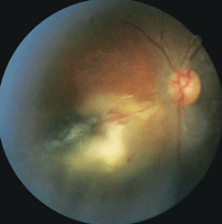
Toxoplasmosis Uveitis and Retinitis
- Most common posterior uveitis and retinitis in immunocompetent individuals
- Ocular inflammation secondary to infection by intracellular parasite.
- Is usually a self-limiting retinochoroiditis, but sight-threatening complications do occur
- Usually 6-8 weeks, with vitreous haze taking a bit longer to clear
- Resolves with atrophic scar and hyperpigmented borders
Risks:
* Immunosuppression and infants, and fatal systemic toxoplasmosis (CNS)
Presentation and Signs:
* Unilateral, sudden floaters, vision loss and photophobia
* Focal retinochoroiditis, affecting posterior pole >50% of the time.
* Necrosis occurs in the inner retinal layer. White fluffy lesions surrounded by retinal oedema.
* Retina is the site for parasite multiplication, but may involve the choroid and sclera
* Secondary anterior uveitis is granulomatous
* Posterior unifocal inflammatory focus near old lesion (satellite lesion)
* MF lesions are uncommon
* Shown in image, severe vitritis (grade) has 'headlight in fog' appearance
* Recurs in 2/3 patients
Other ocular complications:
* Macula involvement has vision effects
* Secondary ONH involvement
* Blood vessel occlusion by inflammatory cells
* Elevated IOP with severe inflammation
Newly Added

CPEO
- Chronic progressive external ophthalmoplegia
- A group of disorders characterised by ptosis and slowly progressive ocular immobility
- Can be isolated, with bilateral ptosis and external ophthalmoplegia
- Can occur in syndromes such as Kearns-Sayrne Sydnrome, Oculopharyngeal dystrophy

Iris Naevus
unfinished
Iris Melanoma
unfinished

Choroidal Melanoma
unfinished
About me

I am Eric Qin, and I am currently the 2025 President of the Giving Sight Society UNSW, studying BVisSci/MClinOptom.
I make this not only for my friends to have fun and practice with, but to raise awareness of the different diseases. This is centred around VISN3111 and OPTM3105 for now.
I try to cover SDGs 3 and 4


Do you have feedback?
I would love more ideas, and any suggestions please put below